


Parts of the Human Ear: Diagram, Functions & NEET Key Points
The concept of structure of ear is essential in biology and helps explain real-world biological processes and exam-level questions effectively. For NEET aspirants, mastering the structure of ear improves accuracy in MCQs and makes revision faster before the exam.
Understanding Structure of Ear
Structure of ear refers to the anatomical organization and function of the human ear, divided into outer ear, middle ear, and inner ear. These parts are important in areas like hearing and equilibrium, human physiology, and sense organs. The structure of ear for NEET includes memorizing names, locations, and functions of each ear part: pinna, auditory canal, tympanic membrane, ossicles (malleus, incus, stapes), eustachian tube, cochlea, vestibule, and semicircular canals. Understanding this concept ensures you can answer diagram-based or function-based questions on the human ear in NEET Biology.
Parts of the Structure of Ear – NEET Essentials
The structure of ear for NEET is split into three major regions, each with unique parts and functions:
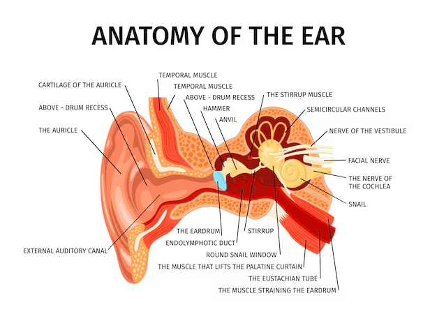
- Outer Ear: Includes the pinna (auricle), external auditory canal, and tympanic membrane (eardrum). Its main job is to collect sound waves and channel them into the ear.
- Middle Ear: Contains the three ossicles (malleus, incus, stapes) and the eustachian tube. The ossicles amplify and transfer sound vibrations, while the eustachian tube equalizes air pressure.
- Inner Ear: Houses the cochlea (for hearing), vestibule, and semicircular canals (for balance).
Each part works in a sequence to convert external sounds into nerve signals and to help maintain body equilibrium. For more, deep dive into the Human Ear page.
Mechanism of Structure of Ear in Hearing
The basic mechanism involves:
- 1. Sound collection: Pinna captures sound waves and sends them through the auditory canal.
- 2. Vibration transmission: Sound waves vibrate the tympanic membrane. These vibrations pass through the malleus, incus, and stapes.
- 3. Signal amplification: Ossicles boost the sound and transmit it to the oval window of the cochlea.
- 4. Conversion to nerve impulse: Inside the cochlea, vibrations become electrical signals in the organ of Corti (hair cells), which are sent to the brain via the auditory nerve.
- 5. Balance: Vestibular apparatus (vestibule and semicircular canals) detects movement for equilibrium.
For a detailed visual, refer to the Labelled Diagram of Human Ear page on Vedantu.
Structure of Ear – Regions, Parts, and Main Functions Table
Here’s a helpful table to understand structure of ear better:
Structure of Ear Table
| Region | Key Parts | Main Function |
|---|---|---|
| Outer Ear | Pinna, Auditory Canal, Tympanic Membrane | Collect sound and channel vibrations |
| Middle Ear | Malleus, Incus, Stapes, Eustachian Tube | Amplify and transmit sound; equalize pressure |
| Inner Ear | Cochlea, Vestibule, Semicircular Canals | Convert vibrations to nerve signals; maintain balance |
Clarifying Key Ear Parts for NEET
- Pinna: Outermost visible part; collects sound
- Tympanic Membrane: Also called eardrum; vibrates due to sound waves
- Malleus, Incus, Stapes: Three small bones (ossicles) in the middle ear; amplify vibrations
- Eustachian Tube: Connects middle ear to throat; balances pressure
- Cochlea: Coiled, snail-like part with sensory hair cells for hearing
- Vestibule, Semicircular Canals: Essential for equilibrium and spatial orientation
Still unclear on the tympanic membrane? Learn more detail at Tympanic Membrane of the Ear or avoid confusion between balance and hearing parts by reviewing Vestibular System.
Common Mistakes to Avoid
- Confusing cochlea (hearing) with vestibule/semi-circular canals (balance).
- Incorrect sequence of ossicles: Always malleus → incus → stapes.
- Forgetting the main job of the eustachian tube (pressure equalization).
- Leaving parts unlabelled in diagrams.
Real-World Applications
The concept of structure of ear is used in fields like ENT medicine (hearing loss, balance disorders), hearing aid design, neuroscience, and audiology. NEET questions often link the ear with neural pathways (see Neurons and Nerves) and with how the brain interprets auditory input (Human Brain page).
For comprehensive sense organ revision, connect ear structure study with Structure of Eye and Structure of Tongue on Vedantu.
In this article, we explored structure of ear, its parts, hearing mechanism, key diagrams, and practical significance for NEET. For maximum confidence, practice more MCQs, revise diagrams, and keep returning to Vedantu’s resources for clear, exam-ready preparation.
FAQs on Structure of Ear – NEET Topic Guide with Diagrams and Functions
1. What is structure of ear in NEET?
The structure of ear in NEET includes three main parts: outer ear, middle ear, and inner ear. The outer ear collects sound vibrations, the middle ear amplifies and transmits these sounds through ossicles (malleus, incus, stapes), and the inner ear contains the cochlea responsible for hearing and the vestibular apparatus for maintaining equilibrium and balance.
2. How to memorise the parts of ear quickly?
To quickly memorise the parts of the ear, use mnemonics and visual aids such as labelled diagrams. For example, remember the ossicles as malleus (hammer), incus (anvil), and stapes (stirrup) by their shapes. Break down the ear into three sections (outer, middle, inner ear) and associate functions with each section. Repeated revision with flashcards and sketching labelled diagrams helps reinforce memory efficiently.
3. What are the three main parts of human ear?
The three main parts of the human ear are:
1. Outer Ear – includes the pinna, external auditory canal, and tympanic membrane.
2. Middle Ear – contains the three ossicles: malleus, incus, and stapes, along with the eustachian tube.
3. Inner Ear – consists of the cochlea responsible for hearing, and the vestibular apparatus that maintains balance.
4. Which ear part maintains balance and equilibrium?
The vestibular apparatus located in the inner ear maintains balance and equilibrium. It includes the utricle, saccule, and three semicircular canals. The utricle and saccule contain maculae with hair cells and otoliths that detect static equilibrium, while the cristae ampullaris in the semicircular canals detect dynamic equilibrium or angular rotation.
5. Are labelled diagrams of the ear important for NEET?
Yes, labelled diagrams of the ear are crucial for NEET preparation. They help in visualising the three parts of the ear, ossicles, cochlea, and vestibular structures which are frequently asked in diagram-based questions and MCQs. Clear diagrams also aid in understanding functions and mechanisms, improving recall during exams.
6. What are the functions of malleus, incus, and stapes?
The ossicles – malleus, incus, and stapes – function to amplify sound vibrations and transmit them from the tympanic membrane to the inner ear. The malleus attaches to the eardrum and passes vibrations to the incus, which then transfers them to the stapes. The stapes connects to the oval window of the cochlea to initiate auditory signal processing.
7. Why do students often confuse the cochlea and vestibule in MCQs?
Students often confuse the cochlea and vestibule because both are part of the inner ear but have distinct functions. The cochlea is responsible for hearing, containing the organ of Corti, while the vestibule is part of the vestibular apparatus responsible for balance. Clear differentiation through diagrams and function-based mnemonics helps avoid this confusion.
8. How can I avoid forgetting the sequence of ear ossicles under exam stress?
To remember the sequence of ear ossicles – malleus, incus, stapes – under exam stress, use simple mnemonics like "MIS" (Malleus, Incus, Stapes) or visualize their shapes: hammer, anvil, stirrup. Practice writing and labelling them repeatedly alongside their function to reinforce retention effectively.
9. What mistakes occur when labelling the ear diagram in NEET?
Common mistakes include misplacing parts like labelling the vestibule as cochlea, confusing the external auditory canal with inner ear structures, or mixing ossicles' names. These errors often stem from unclear memorisation or rushed exam conditions. Consistent practice with clear, labelled diagrams and focus on functional grouping reduces these mistakes.
10. Why is the function of the Eustachian tube commonly misread?
The eustachian tube function is often misunderstood because it does not directly participate in hearing but plays a crucial role in equalising air pressure between the middle ear and atmospheric pressure. This equalisation is essential for proper tympanic membrane vibration. Clarifying that it connects the middle ear to the nasopharynx and controls pressure balance helps avoid this misconception.
11. Are diagram-based questions on ear commonly asked in NEET 2025?
Yes, diagram-based questions on the ear remain a staple in NEET 2025. Test takers are expected to accurately label the outer, middle, and inner ear parts, especially the ossicles, cochlea, and vestibular apparatus. Practising these diagrams helps answer both direct labelling and application-based MCQs confidently.
12. How can I link outer, middle, inner ear to their core functions easily?
Link the three ear parts to their functions as follows:
- Outer ear: collects sound vibrations (pinna and external auditory canal).
- Middle ear: amplifies and transmits sound via ossicles (malleus, incus, stapes) and regulates pressure through the eustachian tube.
- Inner ear: cochlea converts vibrations into neural signals (hearing), vestibular apparatus maintains balance. Associating each part with a simple function mnemonic clarifies their roles quickly.