


Key Differences Between Monocot and Dicot Plant Anatomy for NEET
Understanding the anatomy of monocot and dicot plants is a key part of NEET Biology. This concept helps students differentiate between two major groups of flowering plants by analyzing their internal structures such as roots, stems, and leaves. Accurate knowledge of monocot and dicot anatomy is essential for answering multiple-choice questions, diagram-based queries, and conceptual problems in NEET, making it an important topic for high-scoring preparation.
What is the Anatomy of Monocot and Dicot Plants?
The anatomy of monocot and dicot plants refers to the study of their internal structural organization. Flowering plants (angiosperms) are broadly classified into monocots and dicots based on the number of seed leaves (cotyledons) and various anatomical features visible in their roots, stems, and leaves. Understanding these inner differences helps us explain how plants grow, transport water and nutrients, and perform key life processes.
Core Ideas of Monocot and Dicot Plant Anatomy
Basic Classification: Monocots vs. Dicots
Monocotyledons (monocots) have one seed leaf, while dicotyledons (dicots) have two. This basic difference leads to a series of structural variations throughout the plant body, visible under the microscope and in cross-sections of roots, stems, and leaves. These variations form the foundation for anatomical distinction, crucial in NEET questions.
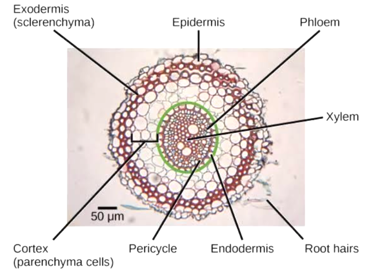
Anatomy of the Dicot Root
Dicot roots typically feature a well-defined cortex, centrally placed stele, and radially arranged vascular bundles. The presence of a large number of xylem and phloem patches and a star-shaped xylem is characteristic. The pericycle is active in forming lateral roots and secondary growth is common.
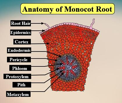
Anatomy of the Monocot Root
Monocot roots have a wide pith, many xylem and phloem patches (polyarch condition), and vascular bundles arranged in a circular manner. Secondary growth is generally absent because the cambium is missing. The number of vascular bundles is usually greater than in dicot roots.
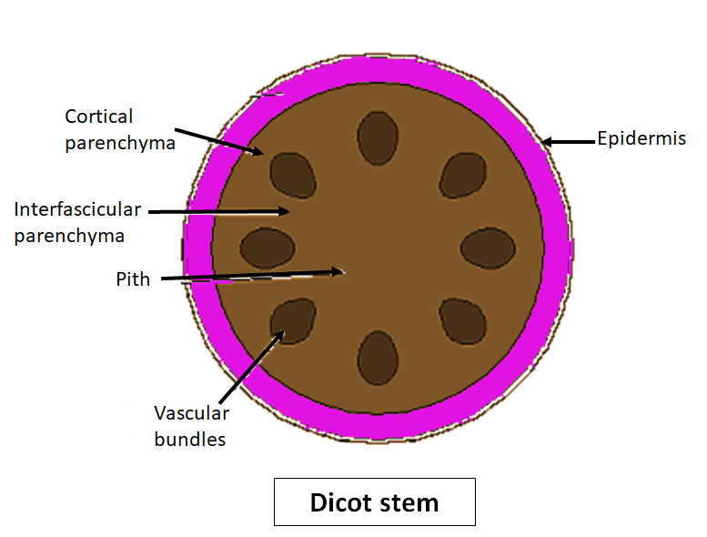
Anatomy of the Dicot Stem
Dicot stems are characterized by vascular bundles arranged in a ring, each bundle being open and collateral with cambium present, which allows for secondary growth. The cortex, endodermis, and pith are well-differentiated. Secondary tissues develop as the plant ages.
Anatomy of the Monocot Stem
Monocot stems have scattered vascular bundles (atactostele) that are closed (no cambium), making secondary growth rare or absent. The bundles are surrounded by a sheath of sclerenchyma. Central pith, cortex, and endodermis may be less distinct.

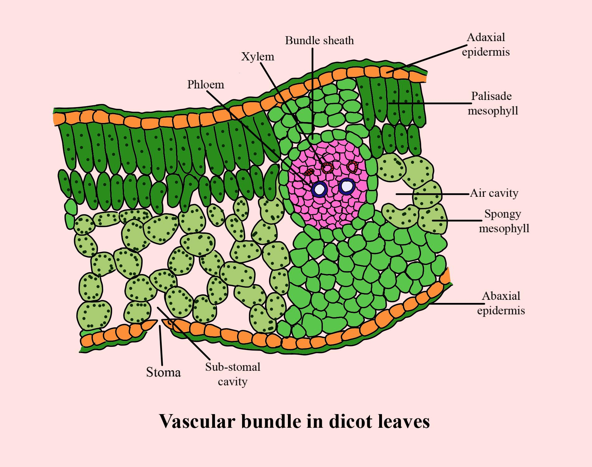
Anatomy of the Dicot Leaf
Dicot leaves display a dorsiventral structure: the upper (adaxial) and lower (abaxial) surfaces differ in anatomy. The mesophyll is differentiated into palisade and spongy parenchyma. Vascular bundles are arranged in a net-like pattern, and the stomata are usually more on the lower surface.
Key Sub-Concepts Connected to Monocot and Dicot Anatomy
Vascular Bundles
Vascular bundles are groups of xylem and phloem tissues responsible for transport. Their arrangement (ringed in dicots, scattered in monocots), type (open or closed), and number (few in dicots, many in monocots) are essential distinguishing features.
Secondary Growth
Secondary growth refers to the increase in thickness of roots and stems due to the activity of the cambium. This is prominent in dicots but generally absent in monocots due to the lack of vascular cambium in monocot stems and roots.
Mesophyll and Leaf Structure
Dicot leaves have a mesophyll differentiated into palisade and spongy layers, supporting efficient photosynthesis. Monocot leaves (not shown here) typically have undifferentiated mesophyll with parallel venation.
Comparative Table: Anatomy of Monocot vs. Dicot Root and Stem
| Feature | Dicot | Monocot |
|---|---|---|
| Number of Vascular Bundles (Stem) | Few, arranged in a ring | Many, scattered |
| Vascular Bundle Type | Open (with cambium) | Closed (without cambium) |
| Secondary Growth | Present | Absent |
| Pith (Root) | Small/absent | Large |
| Arrangement in Root | Radial | Radial |
This table summarizes key differences that help in quick identification. NEET questions frequently involve such comparative features to test a student’s core understanding.
Why is Monocot and Dicot Anatomy Important for NEET?
Questions based on plant anatomy are a recurring part of NEET Biology. Diagrams, comparative tables, and concept-based MCQs test your ability to distinguish between monocot and dicot structures, as well as their functional significance. Mastery of this topic aids in solving questions from plant physiology, development, and genetics, and also helps in interpreting practical botany situations. A deep understanding builds confidence in related chapters like plant tissues, transport, and growth.
How to Study Monocot and Dicot Anatomy Effectively for NEET
- Start by learning the basic classification and fundamental differences.
- Use labeled diagrams to visualize features; practice drawing roots, stems, and leaves of both types.
- Make summary tables for differences and similarities.
- Revise frequently asked MCQs and previous years’ NEET questions.
- Review error patterns, especially in diagram-based questions.
- Correlate anatomy with the functions of each structure for deeper conceptual clarity.
- Practice quick recall of key identification points for exam day.
Common Mistakes Students Make in This Concept
- Confusing stem and root anatomy, especially in vascular bundle arrangements.
- Mixing up concepts of open and closed vascular bundles.
- Forgetting that secondary growth is rare or absent in monocots.
- Mislabeling diagrams or not practicing enough sample diagrams.
- Neglecting the functional significance of anatomical differences.
- Not connecting anatomical features with physiology or other related chapters.
Quick Revision Points: Monocot vs. Dicot Anatomy
- Monocots: one cotyledon, scattered vascular bundles, no secondary growth, large pith in root.
- Dicots: two cotyledons, vascular bundles in rings (stem), open bundles, secondary growth present.
- Dicot root: star-shaped xylem, small pith; Monocot root: circular bundles, large pith.
- Dicot stem: cambium present, permits secondary growth; Monocot stem: cambium absent, secondary growth lacking.
- Dicot leaf: mesophyll differentiated, netted venation; Monocot leaf: generally parallel venation (not shown here).
- Practice diagram labeling and identification for fast recall in exams.
FAQs on Monocot Dicot Plants Anatomy Explained for NEET Students
1. What is the main difference between monocot and dicot plants in NEET biology?
Monocot and dicot plants differ primarily in their seed structure and internal anatomy, which are fundamental NEET exam concepts.
Key differences:
- Monocots have one cotyledon; dicots have two.
- Vascular bundles are scattered in monocots but arranged in a ring in dicots.
- Monocot leaves show parallel venation; dicot leaves have reticulate venation.
- Roots are adventitious in monocots and taproot in dicots.
2. How do you distinguish between monocot and dicot stem anatomy for NEET?
Stem anatomy in monocot and dicot plants can be distinguished by observing the arrangement of vascular bundles.
Differences:
- In monocots, vascular bundles are numerous, scattered, and closed (no cambium).
- In dicots, vascular bundles are fewer, organized in a ring, and open (contain cambium, allowing secondary growth).
3. What are the anatomical features of monocot roots as per NEET syllabus?
Monocot root anatomy is characterized by specific features essential for NEET preparation.
Characteristics:
- Presence of a large, well-developed pith.
- Polyarch xylem (more than six xylem bundles).
- Well-marked endodermis with Casparian strips.
- Vascular bundles arranged in a ring.
4. What is observed in the anatomy of dicot roots?
Dicot root anatomy for NEET is identified by a specific arrangement of tissues.
Key features:
- Prominent cortex and well-developed pericycle.
- Triarch to hexarch xylem (3–6 xylem bundles).
- Small or absent pith.
- Secondary growth is usually present due to open vascular bundles.
5. Why do monocot stems usually not show secondary growth?
Secondary growth is rarely seen in monocot stems because their vascular bundles lack cambium.
Reasons:
- Vascular bundles in monocots are closed (no cambium present).
- Absence of cambium prevents secondary tissue formation and growth in girth.
6. What are the differences between monocot and dicot leaf anatomy for NEET?
The anatomy of monocot vs dicot leaves shows distinct characteristics vital for the NEET exam.
Differences:
- Monocot leaves have parallel venation and similar-sized mesophyll cells (no palisade layer).
- Dicot leaves show reticulate venation and differentiated mesophyll into palisade and spongy parenchyma.
7. What is the significance of endodermis and pericycle in plant root anatomy?
The endodermis and pericycle are key tissues in root anatomy, essential for water regulation and lateral root formation.
Functions:
- Endodermis controls movement of water and minerals into the vascular tissue using Casparian strips.
- Pericycle gives rise to lateral roots and contributes to secondary growth (especially in dicots).
8. How can one identify a monocot root under a microscope?
A monocot root can be identified by several distinct anatomical traits.
Identification tips:
- Large and prominent pith present in the center.
- Polyarch xylem (multiple xylem bundles in a ring).
- Vascular bundles arranged circularly.
- Endodermis with clear Casparian strips.
9. What type of vascular bundle is found in dicot stems?
Dicot stems contain conjoint, collateral, and open vascular bundles, crucial for NEET syllabus.
Details:
- Open vascular bundles have a cambium layer between xylem and phloem.
- This arrangement allows secondary growth (increase in stem thickness) in dicots.
10. Differentiate between hypodermis in monocot and dicot stem.
The hypodermis differs between monocot and dicot stems, an important NEET exam point.
Difference:
- Monocot stem hypodermis is made up of sclerenchyma, giving strength.
- Dicot stem hypodermis is generally made of collenchyma, providing flexibility and support.
11. What are the important points for NEET regarding vascular bundles in monocot and dicot plants?
Vascular bundles are crucial for differentiating monocot and dicot anatomy in NEET biology.
Key points:
- Monocot: Vascular bundles are numerous, scattered, and closed (no cambium).
- Dicot: Vascular bundles are fewer, arranged in a ring pattern, and open (with cambium for secondary growth).
12. Name two monocotyledonous plants and two dicotyledonous plants.
For NEET, it is important to know examples of monocot and dicot plants.
Examples:
- Monocot plants: Maize (Zea mays), Wheat (Triticum aestivum).
- Dicot plants: Pea (Pisum sativum), Sunflower (Helianthus annuus).