

What Are the Main Layers and Functions of the Skin?
The human skin is a multifaceted organ that not only holds our body together but also plays a critical role in shielding us from the outside world. In fact, many experts consider it the largest organ we have, covering roughly 20 square feet in an adult. From regulating temperature to providing our sense of touch, the skin is truly a biological marvel. In this article, we’ll delve into the structure of skin, discuss the 7 layers of skin (including sub-layers), and explore the 10 functions of skin that make it indispensable to our survival. We will also include a handy structure of skin diagram reference to help you visualise these layers more clearly.
Understanding the Structure of Skin
When we talk about the structure of skin, most people refer to three primary layers: Epidermis, Dermis, and Hypodermis (also called the subcutaneous layer). However, you might also come across sources mentioning 7 layers of skin because the outermost epidermis is further divided into multiple sub-layers. Here’s a closer look:
1. Epidermis
The epidermis is the external protective barrier of your body:
Sub-layers of the Epidermis
Stratum Basale (Basal Cell Layer): The deepest sub-layer where new cells (keratinocytes) are produced. Melanocytes (melanin-producing cells) also reside here, determining skin colour and protecting against UV radiation.
Stratum Spinosum (Spinous Layer): Cells here develop spiny structures and start producing more keratin. Langerhans cells, crucial for immune defence, are also found in this region.
Stratum Granulosum (Granular Layer): Keratinocytes in this layer accumulate granules, lose moisture, and gradually die off, preparing for the formation of tougher outer layers.
Stratum Lucidum (Clear Layer): This layer is typically present in thicker areas of skin like the soles of the feet and palms. It appears translucent under the microscope.
Stratum Corneum (Horny Layer): The outermost layer, composed of flattened, dead cells filled with keratin. These cells are tightly packed, forming a waterproof shield.
Together, these five sub-layers in the epidermis are often accounted for when people mention 7 layers of skin (the remaining two being the dermis and hypodermis).
2. Dermis
Beneath the epidermis, the dermis offers strength and flexibility:
Dermal Papillae: Finger-like projections that increase the surface area between the dermis and epidermis, aiding the supply of nutrients.
Collagen and Elastin Fibres: Provide structural support, elasticity, and strength.
Blood Vessels: Supply essential nutrients and oxygen to the skin cells and play a crucial role in temperature regulation.
Nerves: Detect touch, temperature, pressure, and pain, transmitting signals back to the brain.
Hair Follicles: Tubular structures anchoring hair strands. Each hair follicle is attached to a tiny arrector pili muscle that contracts to produce “goosebumps.”
Sebaceous (Oil) Glands: Produce sebum, a natural lubricant for the skin that helps guard against microbes and keeps the skin hydrated.
Sweat Glands: Found all over the skin; they release sweat to regulate body temperature and excrete minor metabolic waste.
3. Hypodermis (Subcutaneous Layer)
Often overshadowed by the epidermis and dermis, the hypodermis is a vital layer:
Adipose (Fat) Tissue: Acts as an insulator against both heat and cold, provides an energy reserve, and cushions internal organs from mechanical shocks.
Larger Blood Vessels and Nerves: Support deeper circulation and sensation.
Thickness Variations: The thickness of this layer varies greatly depending on age, health, and body region. Around the eye region, for instance, it’s thinner to allow free movement of the eyeball.
Explore, Human Body System
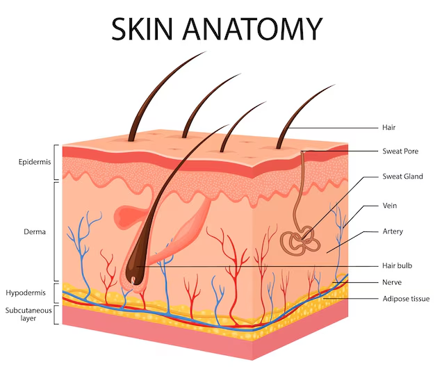
Structure of Skin Diagram
A structure of skin diagram can be highly beneficial for seeing how each layer stacks up. Look for a labelled illustration that distinctly shows the epidermal sub-layers (especially the Stratum Lucidum in thicker regions), the dermis with its hair follicles and glands, and the hypodermis rich in fat cells. Observing such a diagram helps you appreciate the complexity and arrangement of tissues in your body’s protective shield.
10 Functions of Skin
While many textbooks condense the function of skin into a few key roles, let’s explore the 10 functions of skin that encompass everything from protection to communication:
Protection from Pathogens: The skin forms the first line of defence, preventing harmful microbes from penetrating deeper tissues.
Barrier Against External Elements: Thickened layers, especially in areas like soles and palms, prevent mechanical and chemical damage.
Prevents Excessive Water Loss: A healthy skin barrier retains moisture, crucial in arid climates where dehydration risk is higher.
Temperature Regulation: Sweating and blood vessel dilation in the dermis help cool the body, while constriction helps retain heat.
Sensory Reception: Millions of nerve endings detect touch, pain, pressure, temperature changes, and more, ensuring appropriate responses to the environment.
Vitamin D Synthesis: Upon exposure to sunlight, specific cells in the epidermis use UV rays to start vitamin D production, essential for calcium absorption and bone health.
Excretion of Wastes: Sweat glands help flush out minor amounts of urea, salt, and other metabolic by-products, maintaining internal balance.
Camouflage and Colouration (in Some Animals): While humans don’t actively camouflage, variations in melanin production offer colour adaptation and UV protection. Certain animals can alter their skin patterns to blend with surroundings or communicate.
Fat Storage and Shock Absorption: The hypodermis stores adipose tissue that acts as an energy reserve and a cushion against impacts.
Chemical Signalling: The sweat and oils on our skin can contain pheromones and unique scents, sending signals to others (more pronounced in animals).
Explore: Difference Between Epidermis and Dermis
Tips for Healthy Skin
Beyond the standard structure of skin, there are fascinating aspects that many people overlook:
Skin pH and Microbiome: A slightly acidic pH helps deter harmful bacteria, while nurturing a healthy population of beneficial microbes that live on the skin’s surface.
Role of Hydration: Adequate water intake and balanced nutrition support the epidermal barrier, preventing dryness and flaking.
Sun Protection: Overexposure to UV light can damage the dermis and increase the risk of skin cancers. Sunscreen and protective clothing are essential.
Also, read Sense Organs
Interactive Quiz: Test Your Skin Knowledge
Which layer of the skin contains melanocytes?
A. Stratum Corneum
B. Stratum Basale
C. Granular Layer
D. Spinous Layer
Which term refers to the subcutaneous layer of the skin?
A. Epidermis
B. Dermis
C. Hypodermis
D. Stratum Lucidum
Which of the following is NOT a primary function of skin?
A. Temperature Regulation
B. Protection from UV Rays
C. Producing Digestive Enzymes
D. Sensory Reception
Which protein is predominantly found in hair, nails, and the outer skin cells?
A. Keratin
B. Melanin
C. Collagen
D. Elastin
Which layer is responsible for binding the epidermis and dermis?
A. Basement Membrane (Dermo-Epidermal Junction)
B. Stratum Lucidum
C. Dermal Papillae
D. Granular Layer
Check Your Answers Below
B. Stratum Basale
C. Hypodermis
C. Producing Digestive Enzymes
A. Keratin
A. Basement Membrane (Dermo-Epidermal Junction)


FAQs on Structure and Functions of Human Skin: Complete Student Guide
1. What are the principal functions of the human skin?
The human skin performs several vital functions essential for survival. Its primary roles include:
- Protection: It acts as a physical barrier against pathogens, UV radiation, chemical damage, and dehydration.
- Thermoregulation: It maintains body temperature through sweating and regulating blood flow.
- Sensation: It houses numerous receptors for touch, pressure, pain, and temperature.
- Excretion: It eliminates waste products like urea and salts through sweat.
- Vitamin D Synthesis: It synthesizes Vitamin D when exposed to sunlight, which is crucial for bone health.
2. What are the three main layers of the skin and their sub-layers?
The skin is composed of three primary layers:
1. The Epidermis: The outermost, avascular layer responsible for protection. It has five sub-layers: Stratum Corneum, Stratum Lucidum, Stratum Granulosum, Stratum Spinosum, and Stratum Basale.
2. The Dermis: The middle layer containing blood vessels, nerves, glands, and hair follicles.
3. The Hypodermis (Subcutaneous Layer): The deepest layer, composed of fat and connective tissue, which provides insulation and anchors the skin to underlying muscles.
3. How do the epidermis and dermis differ in their structure and function?
The epidermis and dermis differ significantly. The epidermis is the thin, outer layer made of epithelial tissue and is avascular (lacks blood vessels); its main function is to provide a protective barrier. In contrast, the dermis is the thicker, inner layer made of dense connective tissue. It is highly vascularised and contains nerves, glands, and hair follicles, providing structural support, sensation, and nourishment to the epidermis.
4. Can you describe the key structures found within the dermis layer?
The dermis is a complex layer that houses many essential structures. Key components include:
- Blood Vessels: To nourish the skin and regulate temperature.
- Nerve Endings and Receptors: For detecting touch, pressure, pain, and heat.
- Hair Follicles: Sheaths of cells and connective tissue that surround the root of a hair.
- Glands: Including sebaceous (oil) glands and sudoriferous (sweat) glands.
- Collagen and Elastin Fibres: Proteins that provide the skin with strength and elasticity.
5. What is the importance of melanin in the skin?
Melanin is a pigment produced by cells called melanocytes in the epidermis. Its primary importance is to protect the skin from harmful ultraviolet (UV) radiation from the sun. It absorbs UV rays, preventing them from damaging the DNA in skin cells, which could otherwise lead to skin cancer. Melanin is also the main determinant of skin and hair colour.
6. Why is the skin on the palms and soles thicker than on other parts of the body?
The skin on the palms of the hands and soles of the feet is thicker primarily as a functional adaptation to high friction and mechanical stress. This thickness is due to a significantly more developed stratum corneum (the outermost epidermal layer). This type of skin, known as 'thick skin', also contains an extra layer called the stratum lucidum, which is absent in 'thin skin' found elsewhere on the body.
7. How does the skin function as a major sensory organ?
The skin functions as a major sensory organ because it is densely packed with various types of specialised nerve endings and receptors, primarily in the dermis. These receptors detect different stimuli from the external environment. For example, Meissner's corpuscles detect light touch, Pacinian corpuscles detect pressure and vibration, while free nerve endings sense pain and temperature changes, relaying this information to the nervous system.
8. What are the different types of glands in the skin and what are their examples and roles?
The skin contains two main types of exocrine glands:
1. Sebaceous Glands: These are oil glands, typically attached to hair follicles. They secrete an oily substance called sebum, which lubricates the hair and skin, prevents water loss, and has antimicrobial properties.
2. Sudoriferous Glands: These are sweat glands responsible for producing sweat. There are two types: eccrine glands, which are found all over the body and help in thermoregulation, and apocrine glands, found in areas like the armpits and groin, which produce a thicker secretion.
9. How does the skin repair itself after being damaged, such as from a cut?
The skin's repair process depends on the depth of the injury. For a superficial cut affecting only the epidermis, cells from the stratum basale divide and migrate to fill the gap. For deeper cuts involving the dermis, a more complex process occurs: an inflammatory response cleans the wound, followed by the formation of granulation tissue. Fibroblasts then produce collagen fibres to close the wound, which may result in the formation of scar tissue that is structurally different from the original skin.
10. What is a key medical application that relies on a deep understanding of the skin's structure?
A key medical application is transdermal drug delivery. By understanding the structure and permeability of the skin's layers, especially the barrier function of the stratum corneum, medical science has developed medicated patches. These patches deliver a controlled dose of medication (such as hormones, nicotine, or pain relievers) through the skin directly into the bloodstream over an extended period, which can improve patient compliance and reduce side effects associated with oral administration.


