


An Overview of Class 9 Biology Study The Structure Of Plant And Animal Tissues And Draw Their Well Labelled Diagrams Experiment
Biology experiment - Studying the Structure of Plant and Animal Tissues and Drawing their Well-labelled Diagrams
Cells are the structural and functional unit of life. A cell is the smallest unit of the living body which can carry out all life processes, such as, nutrition, excretion, respiration, etc. Similar structured cells coming together, to perform similar functions, are termed as tissues. As we know that plant and animal cells are different in structure; tissues possessed by plants and animals are also different. Plant tissues are of various types, such as meristematic, permanent, epidermal tissues, etc. Animal tissues are divided into four types, epithelial, connective, nervous and muscular, based on the type of function they are performing.
Table of Contents
Aim
Apparatus required
Theory
Procedure
Observation
Result
Precautions
Aim
To study the structure of different types of animal, plant tissues and draw well labelled diagrams.
Materials required
Methylene blue stain, slides of nervous tissue, muscular tissue, collenchyma, parenchyma and sclerenchyma, compound microscope, etc.
Theory
Classification of Plant tissue
Depending on ability to divide plant tissues are divided into Meristematic and permanent tissues.Mestimtimatic tissues have power of cell division throughout the life of the plant.Permanent tissues lose the power of cell division and attain a definite shape and size and perform a definite function. Plant tissues are further differentiated as given in the diagram.
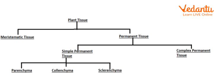
Flow chart of Plant tissue
Classification of Animal Tissu
Group of cells having the same origin and performing the same tasks are known as tissues. During the embryonic development of higher animals there are 3 germ layers- Ectoderm, Mesoderm and Endoderm. These germ layers differentiate and lead to formation of various types of cells and tissues. Animal tissues are further divided as given below in the diagram-
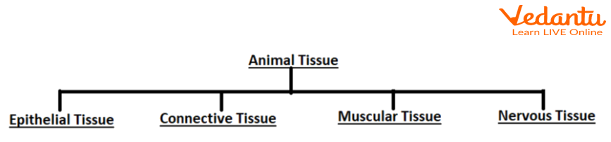
Flow chart of Animal Tissue.
Procedure
Carefully take some clean cotton and rub it on the inner side of cheeks. Tap the cotton on the clean glass slide and put one drop of blue stain on it and cover with coverslip.
Carefully take out the prepared slides of Collenchyma, sclerenchyma, parenchyma, Muscular tissue and nervous tissue.
Observe the structure of tissues in a compound microscope and draw well labelled diagrams.
Observation
Parenchyma-
Parenchyma cells are isodiametric.
Intercellular space can be observed.
Possess large central vacuole.
Peripheral cytoplasm with a nucleus is present.
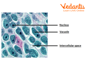
Parenchyma tissue
Collenchyma-
The cells are kind of oval or elongated.
Cells are thickened at the corners.
Intercellular spaces are not present.
Cells possess large vacuoles and prominent nuclei.
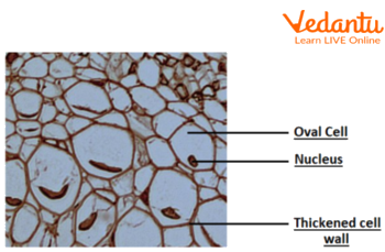
Collenchyma tissue
Sclerenchyma-
Sclerenchyma cells are dead.
Pits are present acting as connections with adjacent cells.
Thickened cell walls consist of lignin.
They provide support and mechanical strength to the plant.
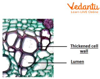
Sclerenchyma tissue
Cheek cell-
Proper nucleus is visible.
Irregular shaped cell.
Cells with cell membranes are present.
Very less intercellular space is seen.
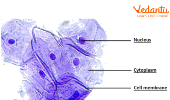
Cheek Cell
Muscular Tissue-
Skeletal Muscles-
Multinucleated cells.
These muscles are attached to the skeleton of the body.
These muscle cells are cylindrical, elongated and enclosed in a membrane called sarcolemma.
These muscles possess light and dark bands which give it stripped appearance, giving them another name as striated muscle.
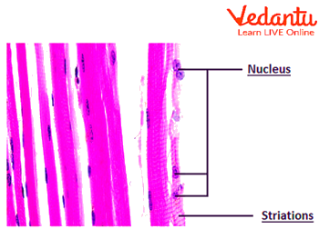
Skeletal muscle
Smooth muscle-
Spindle shaped cell.
Centrally located nucleus is present.
These cells do not possess striations.
Commonly found in the alimentary canal and blood vessels.
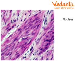
Smooth muscle
Cardiac muscle-
They are long, branched and uninucleated cells.
They show striations that are dark and light bands.
Presence of intercalated discs.
Cardiac muscles are present only in the walls of the heart.
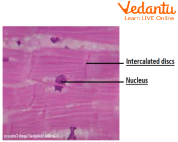
Cardiac Muscle
Nerve cell-
Long cytoplasmic projection arises from a cell body known as an axon.
Myelin sheath is present over the axon in some nerve fibres.
Branched projections arise from the cell body which are termed as Dendron.
Nerve cells consist of a cell body with a single nucleus and cytoplasm.
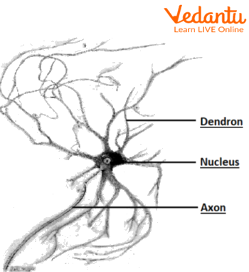
Nerve cell
Result
Precautions
Make sure to stain the cells properly
Adjust the compound microscope well.
Lab Manual Questions
1. What type of cells are possessed by sclerenchyma?
Ans: Sclerenchyma possesses dead cells. It contains a thick layer of lignin and it provides mechanical support to the plant and protects the inner parts of the plant.
2. Explain the features of striated muscle fibre.
Ans: The striated muscle fibres are multinucleated, unbranched and cylindrical. They are striated, because of the presence of dark and light bands.They are found attached to the skeletal bones.
3. State the difference between nervous tissue and epithelial tissue.
Ans: The epithelial tissue consists of irregular simple cells with a nucleus whereas the nervous tissues possess the nerve cell which have extensions like axon and dendron which helps in easy identification.
4. How can you differentiate between a parenchyma and collenchyma slide?
Ans: The parenchyma cells are isodiametric but the collenchyma cells are not. The collenchyma cells have thick cell wall corners whereas the parenchyma cells have thin cell walls.
Viva Questions
1. Define meristematic tissues.
Ans: The cells which are capable of dividing throughout their life are known as meristematic cells.
2. What is differentiation?
Ans: The process by which cells lose their property of division and are assigned specialised functions is termed as differentiation.
3. Define permanent tissues and name the types of permanent tissues.
Ans: The cells which lose their ability to divide and perform special functions are known as permanent tissues.
There are two types of permanent tissues- Simple and complex.
4. Explain simple tissues with examples.
Ans: Simple tissue is a type of permanent tissue. They possess groups of similar types of cell performing similar functions.
Examples of simple tissues are- Parenchyma, collenchyma and sclerenchyma.
5. Explain complex tissue with examples.
Ans: Complex tissue is a type of permanent tissue possessing different types of cells performing similar functions.
Examples of complex tissue are- Xylem and phloem.
6. What do you mean by histology?
Ans: Histology is the branch of biology which deals with studies of tissues.
7. Which type of animal tissue possess intercalated discs?
Ans: Intercalated discs are possessed by cardiac muscle tissue.
8. Name the longest cell in an animal body.
Ans: Nerve cell is the longest cell in the animal body.
9. Which part of the cell gives rise to axon and dendron?
Ans: The axon and dendron arise from the cell body or cyton.
10. Which type of animal tissue possesses the light and dark band?
Ans: The skeletal muscle tissue and the cardiac muscle tissue possesses the light and dark band giving them the prominent striations.
Practical Based Questions
1. The cell with tapering end and elongated body structure-
Sclerenchyma
Nerve cell
Parenchyma
Smooth muscles
Ans: Smooth muscle
2. The long tube like projection originating from the cyton is-
Cell body
Axon
Dendron
Myelin sheath
Ans: Axon
3. A student can identify a skeletal muscle because of-
Striations absent
Striations present
Striations present and multinucleated
Striations are present and multinucleated.
Ans: Striations present and multinucleated
4. Smooth muscle fibres are-
Spindle shaped, striated, and involuntary.
Cylindrical, unbranched, striated, multinucleate and involuntary.
Spindle shaped, non-striated, and involuntary.
Cylindrical, striated, unbranched, multinucleate and voluntary.
Ans: Spindle shaped, non-striated, and involuntary
5. The stain used for staining the slides is-
Safranin
Iodine
Methylene blue
Acetocarmine
Ans: Methylene blue
6. If you are given slides of parenchyma and sclerenchyma, you will identify sclerenchyma based on-
Thickness of cell wall
Presence of sarcolemma
Location of nucleus
Position of vacuoles
Ans: Thickness of cell wall
7. The cells which actively divide comes under the category of-
Collenchyma
Meristematic
Permanent
Parenchyma
Ans: Meristematic
8. Which of the following possesses a thin wall?
Parenchyma
Sclerenchyma
Collenchyma
All of these
Ans: Parenchyma
9. Which of the following will have thick cell walls and is non-living?
Collenchyma
Parenchyma
Complex tissue
Sclerenchyma
Ans: Sclerenchyma
10. The cheek cells comes under which type of animal tissue-
Striated muscle
Unstriated muscle
Nerve cell
Epithelial tissue
Ans: Epithelial tissue
Summary
Depending on their ability of division, plant tissues are divided into two categories- Meristematic and permanent. Permanent tissues lose their ability to divide and meristematic tissues can undergo cell division through their entire life.The animal tissue depending where they are found and their function, they are divided into 4 categories- Epithelial, muscular, connective and nervous.


FAQs on Class 9 Biology Study The Structure Of Plant And Animal Tissues And Draw Their Well Labelled Diagrams Experiment
1. What are the most important differences between plant and animal tissues that students must know for the Class 9 exam?
For the CBSE Class 9 exam, the key differences between plant and animal tissues are crucial. Students should focus on the following points:
- Mobility: Plant tissues are largely supportive and stationary, providing structural strength. Animal tissues are adapted for mobility, enabling movement.
- Growth: Growth in plants is restricted to specific regions with meristematic tissue. In animals, cell growth is more uniform throughout the body.
- Structural Organisation: The organisation of plant tissues is simpler compared to the highly specialised and complex organ systems found in animals.
- Energy Needs: Plants have more dead supportive tissues, leading to lower energy requirements. Animals have mostly living tissues and require more energy for locomotion and other life processes.
2. Which diagram is frequently asked from the animal tissues section for 5 marks?
One of the most important diagrams for a 5-mark question is that of a neuron or nerve cell. A well-labelled diagram should include the following parts:
- Cyton (Cell Body): Contains the nucleus and cytoplasm.
- Dendrites: Short, branched fibres that receive signals.
- Axon: A single, long fibre that transmits signals away from the cell body.
- Myelin Sheath: An insulating layer covering the axon (optional but good to show).
- Nerve Ending: The terminal part of the axon.
3. Why is blood considered a type of connective tissue? This is a common HOTS question.
Blood is considered a connective tissue because, like other connective tissues, it has two main components: cells and an extensive extracellular matrix. However, its matrix is fluid.
- The Matrix: The fluid matrix of blood is called plasma. It contains water, proteins, salts, and hormones.
- The Cells: Suspended within the plasma are the living cells: Red Blood Cells (RBCs), White Blood Cells (WBCs), and platelets.
4. How can you differentiate between the three types of simple permanent tissues in plants for a 3-mark question?
To differentiate between parenchyma, collenchyma, and sclerenchyma, focus on these three key aspects:
- Parenchyma: Composed of living cells with thin cell walls and large intercellular spaces. Its main functions are storage of food and providing support.
- Collenchyma: Composed of living cells that are irregularly thickened at the corners, with very little intercellular space. It provides mechanical support and flexibility to plant parts like stems and leaves.
- Sclerenchyma: Composed of dead, long, and narrow cells. Their walls are heavily thickened with lignin, leaving no internal space. Its sole purpose is to provide hardness and stiffness to the plant.
5. What are the three types of muscle tissues found in animals? State one key feature and location for each.
The three types of muscle tissues are:
- Striated Muscles (Skeletal Muscles): These are voluntary muscles with light and dark bands (striations). They are attached to bones and help in body movement. Example: Muscles of the limbs.
- Smooth Muscles (Unstriated Muscles): These are involuntary muscles with spindle-shaped cells and no striations. They are found in the walls of internal organs. Example: Muscles in the alimentary canal.
- Cardiac Muscles: These are involuntary muscles found only in the heart. They are cylindrical, branched, and show striations. They contract rhythmically throughout life.
6. What is the importance of complex permanent tissues in plants?
Complex permanent tissues are crucial for the survival of land plants as they form the vascular or conducting system. They are made of more than one type of cell that work together.
- Xylem: It conducts water and minerals from the roots to other parts of the plant. It consists of tracheids, vessels, xylem parenchyma, and xylem fibres. It also provides mechanical strength.
- Phloem: It transports food (prepared during photosynthesis) from the leaves to storage organs and other parts of the plant. It consists of sieve tubes, companion cells, phloem parenchyma, and phloem fibres.
7. Why do students often confuse tendons and ligaments? What is the main difference an examiner looks for?
Students confuse tendons and ligaments because both are types of dense connective tissue involved in the skeletal system. The key difference an examiner looks for is what they connect:
- Tendons: Connect muscle to bone. They are fibrous, have great strength, but limited flexibility.
- Ligaments: Connect bone to bone. They are highly elastic and flexible, providing stability to joints.
8. What is the role of the epidermis in plants and how is it adapted for protection?
The epidermis is a protective tissue that forms the outermost layer of the entire plant body. Its primary role is protection against water loss, mechanical injury, and invasion by parasitic fungi. In desert plants, the epidermis has special adaptations, such as a thick, waxy coating of cutin on its surface, which is a chemical substance that makes it waterproof and reduces the rate of transpiration.
9. From an exam perspective, what are meristematic tissues and their types based on location?
Meristematic tissues are plant tissues containing actively dividing cells responsible for plant growth. For exam purposes, it's important to know their types based on location:
- Apical Meristem: Located at the tips of roots and shoots, it is responsible for the increase in the length of the plant.
- Lateral Meristem (Cambium): Found along the sides of stems and roots, it is responsible for the increase in the girth or width of the plant.
- Intercalary Meristem: Located at the base of leaves or internodes, it helps in the longitudinal growth of organs.
10. Cardiac muscles are involuntary, so why do they have striations like voluntary skeletal muscles?
This is an excellent higher-order thinking question. Cardiac muscles have striations (light and dark bands) due to the regular arrangement of contractile proteins (actin and myosin), similar to skeletal muscles. This arrangement allows for powerful, coordinated contractions necessary to pump blood effectively. However, their function is involuntary because their contraction is not under conscious control. The presence of intercalated discs, which are unique to cardiac muscle, allows for the rapid spread of electrochemical signals, ensuring the heart contracts as a single, synchronised unit.