


An Overview of Class 12 Biology Mitosis And Its Different Phases In A Temporary Mount Of An Onion Root Tip Experiment
Do you know how plant cells are able to divide and grow? Mitosis is the process which helps them to keep on dividing. Do you know which cells in the pants are capable of dividing themselves? In plant cells, cells divided by mitosis take place in meristematic tissues (called meristems). The cells in meristems are undifferentiated and are formed at the growing tip of roots and shoots and in the cambium between the xylem and phloem.
To know more about this experiment continue reading this article.
Table of Content
Aim
Apparatus required
Theory
Procedure
Observation
Result
Precautions
Lab Manual Questions
Viva Questions
Practical Based Questions
Conclusion
FAQs
Aim
To study mitosis by preparing a temporary mount of an onion root tip.
Apparatus Required
Onion
Glass slide
Filter paper
Aceto-alcohol
Cover slip
N/10 hydrochloric acid
Acetocarmine stain
Water
Watch glass
Forceps
Blade
Dropper
Needle
Burner
Compound microscope
Theory
Somatic cells divide through equational division that allows them to divide and produce their daughter cells wherein the number of chromosomes remains the same (i.e., unchanged) as in the original cell. In plants, these divisions occur in meristematic tissues of roots and shoot tips as all the stages of mitosis are clearly observable.
Procedure
The first step is to grow root tips of onion bulbs:
Carefully remove the dry roots present on medium-sized onion bulbs.
Place the onion bulbs on glass tubes filled with water in such a way that the stem portion of the bulb just touches the water.
Cut 2-3 cm long freshly grown roots and transfer them to freshly prepared aceto-alcohol.
Aceto-alcohol is prepared by taking glacial acetic acid and ethanol in the ratio of 1:3.
Keep the root tips in the fixative for nearly 24 hours and then transfer them to 70% ethanol.
Onion root tip cells divide once in 24 hours and this division usually takes place 2 hours after sunrise which is why root tips grown in water should be cut at that time only to get the maximum number of dividing cells.
Now, the following steps should be done to prepare the glass slide.
Wash one or two preserved roots in water and place them in a clean and grease free slide.
Place one drop of N/10 hydrochloric acid on the root tip and then 2-3 drops of aceto-carmine stain on it.
Place the glass slide over a spirit lamp and warm it for 5-10 minutes. (Note:- Be careful while warming up the slide so that the stain should not dry up).
Blot the excess of stain using blotting paper and retain a comparatively more stained portion of root tip and discard the remaining portion.
Now after 10 seconds put 1-2 drops of water and blot the excess water using blotting paper and mount the cover slip carefully over the root tip to avoid air bubbles.
Using the blunt end of a pencil, slightly tap the cover slip mounted so that the meristematic tissues are properly squashed and spread as a thin layer of cells.
The following steps describe how to study the prepared slide.
Place the slide under the compound microscope and first study the slide under lower magnification to search the areas of dividing cells.
Now examine the slide under higher magnification to observe the detailed phases of mitosis.
Observation
Karyokinesis and cytokinesis are the two stages of mitosis and the cells which are not in the dividing phase are said to be in the interphase.
Interphase:-
In this phase the cell prepares itself for the cell division by undergoing both cell growth and DNA replication in a sequential manner. The chromatin material looks granular and the boundary of the nucleus is distinct and clearly observable.
Stages of Mitosis:-
Prophase- During prophase condensation of chromatin material takes place and centrioles will start moving towards the opposite poles. The nuclear membrane disintegrates and cell organelles like endoplasmic reticulum, nucleolus and Golgi complex disappear. Centrosomes move towards the opposite pole and each centrosome radiates out microtubules called asters. Two aster together with spindle fibres form mitotic apparatus.
Metaphase- The condensation of chromosomes is completely done and are clearly observed under a microscope. The spindle fibres get attached to the kinetochores of chromosomes. The chromosomes align themselves at the equator of the cell forming a metaphase plate.
Anaphase- During this stage, each chromosome is split simultaneously and the two daughter chromatids start moving towards the opposite poles. The centromere of each chromosome is facing towards the pole while the arms of the chromosomes are trailing behind.
Telophase- By this stage the chromosomes have reached their respective poles and decondense and lose their individuality. The chromosomes appear to be a mass at the two poles. The cell organelles also reform.
Cytokinesis
In plants, the division of cytoplasm starts in the centre of the cell and grows outwards to meet the existing lateral walls and ultimately divides the cell into two. Whereas in animal cells the cytokinesis starts by the appearance of a furrow in the plasma membrane and deepens and ultimately joins at the centre that divides the cell into two. In some cells cytokinesis is not followed by karyokinesis which raises the multinucleated condition leading to the formation of syncytium.
Result
The different stages of mitosis in an onion cell are observed under the microscope.
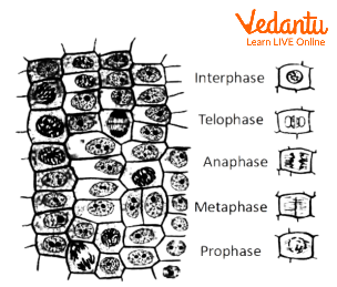
The different stages of mitosis in an onion cell
Precautions
Take all the chemicals in a minimal amount to avoid wastage.
Remove excess water and stain using a blotting paper and make a clean slide to observe under the microscope.
Be careful while warming up the glass slide so that the stain does not dry up.
Observe the slide under lower magnification to find the areas of dividing cells.
Place the coverslip very carefully without any air bubbles.
Lab Manual Questions
1. Suggest names of a few tissues which are suitable for the study of mitosis?
Ans: As root tips have meristematic tissues which divide rapidly so the study of mitosis can be easy. Thus, root tips of onion, wheat, barley, lentils etc. can be used for the study of mitosis.
2. Why is mitosis also called equational division?
Ans: Mitosis is also called equational division as at the end of each division, each new cell has the same number of chromosomes as in the original cell.
3. What shape would a metacentric and submetacentric chromosome exhibit during the anaphase stage?
Ans: The metacentric chromosomes appear to be V-shaped whereas the sub-metacentric chromosomes appear to be L-shaped during the anaphase stage.
4. How does cytokinesis differ in plant and animal cells?
Ans: Cytokinesis is the division of cytoplasm. In plant cells, cytokinesis starts from the middle of the cytoplasm and grows outwards while in an animal cell, the cytokinesis starts due to the appearance of a furrow in the plasma membrane that grows towards the centre of the cell ultimately dividing the cell.
Viva Questions
1. What are the two main stages of mitosis?
Ans: The two main stages of mitosis are cytokinesis and Karyokinesis.
2. What is the most suitable material for study of different phases of mitosis?
Ans: Root tips are the most suitable material for study of different phases of mitosis.
3. What are the different stages of interphase?
Ans: The different stages of Interphase are G1 (Gap 1) phase, G2 (Gap 2) phase and the synthesis phase (S phase).
4. At what stage does the cell spend most of its life?
Ans: The cell spends most of its life in the interphase stage.
5. In which phase does the disintegration of cell organelles start and end?
Ans: The disintegration of cell organelles start by the starting of early prophase and is completed by the end of late prophase.
6. What are kinetochores?
Ans: The kinetochores are the specialised disc-shaped structures around centromere and it allows the attachment of spindle fibres to the chromosomes.
7. Which structure pulls the chromatids towards the opposite poles?
Ans: The spindles fibres pull the chromatids towards the opposite pole.
8. What is the characteristic feature of metaphase?
Ans: The metaphase plate is the characteristic feature of metaphase where the chromosomes align themselves at the equator of the cell.
9. By what does the Go phase is characterised?
Ans: There is no cell division in the Go phase and is thus known as the resting phase.
10. What is the first stage of karyokinesis?
Ans: The first stage of Karyokinesis is the prophase.
Practical Based Questions
Chemicals required to make acetocarmine stain?
Glacial acetic acid and ethanol
Ethanol and glycerine
Glacial acetic acid and methanol
DAPI
Answer:- A
What other stain can be used in place of aceto-carmine?
Methylene blue
Safranin
Aceto-orcein
Fast green
Answer:- C
The two main stages of cell cycle are:-
Cytokinesis and Interphase
Prophase and Metaphase
Telophase and Karyokinesis
Karyokinesis and Cytokinesis
Answer:- D
Which of the following is the best material for the study of mitosis?
The sclerenchyma cells in the stem.
The meristematic cells present in the root tip.
Xylem cells.
The cells of the leaf.
Answer:- B
Is acetocarmine acidic or basic?
Highly acidic
Highly basic
Mildly acidic
Mildly basic
Answer:- C
Which structure pulls the chromatids to the opposite poles of the cell?
Spindle fibres
Chromatin fibres
Kinetochores
Centromere
Answer:- A
Which stage of mitosis is longest?
Prophase.
Telophase
Metaphase
Anaphase
Answer:- A
What stage of mitosis is characterised by the presence of an equatorial plate?
Metaphase
Prophase
Telophase
Anaphase
Answer:- A
Conclusion
In this article, we have studied an experiment on mitosis in an onion root tip.
We have seen the different stages of mitosis.
These stages have different characteristics and are collectively termed as karyokinesis. The cytokinesis is followed by the karyokinesis.
We have also seen certain precautions taken while carrying out the procedure.
FAQs on Class 12 Biology Mitosis And Its Different Phases In A Temporary Mount Of An Onion Root Tip Experiment
1. What kind of short, 1-mark questions are frequently asked from the Mitosis topic in the CBSE Class 12 board exams?
For 1-mark questions, you can expect direct queries about specific stages or events. For the 2025-26 exams, focus on questions like:
- Which stage of mitosis is best for studying the morphology of chromosomes? (Answer: Metaphase)
- In which stage of the cell cycle do cells that do not divide further exit? (Answer: They enter the quiescent stage or G0)
- What is the main function of the mitotic spindle? (Answer: To separate sister chromatids during Anaphase)
2. How should I structure a 3-mark answer explaining the significance of mitosis in living organisms?
To secure full marks, you should clearly list at least three distinct points. A good answer would include:
- Growth and Development: Mitosis is the primary reason for the growth of multicellular organisms from a single-celled zygote into a complex adult body.
- Cell Replacement and Repair: It continuously replaces old or damaged cells, such as skin cells, gut lining cells, and red blood cells, helping in tissue repair and maintenance.
- Asexual Reproduction: In many unicellular organisms like amoeba and in the vegetative propagation of plants, mitosis is the method of reproduction.
3. What are the key differences between mitosis and meiosis that are most important for the board exam?
This is a very important topic. For your exam, focus on these key distinctions:
- Type of Division: Mitosis is an equational division, meaning the chromosome number in daughter cells is the same as the parent cell. Meiosis is a reductional division, where the chromosome number is halved.
- Number of Divisions: Mitosis is completed in one nuclear division, while meiosis involves two successive divisions (Meiosis I and Meiosis II).
- Genetic Variation: Mitosis produces genetically identical daughter cells. Meiosis introduces genetic variation through processes like crossing over.
- Purpose: Mitosis occurs for growth and repair in somatic cells, while meiosis occurs in germ cells to produce gametes for sexual reproduction.
4. How can I identify the different stages of mitosis in an onion root tip cell diagram, a common exam question?
To identify the stages in an onion root tip cell, look for these specific visual cues:
- Prophase: You'll see condensed, thread-like chromosomes scattered inside the nucleus. The nuclear membrane is usually still intact.
- Metaphase: This is the easiest to spot. The chromosomes are thick, clearly visible, and aligned in a straight line at the cell's equator, known as the metaphase plate.
- Anaphase: Look for sister chromatids being pulled apart towards opposite ends (poles) of the cell. They often appear V-shaped or J-shaped.
- Telophase: Two distinct clusters of chromosomes are present at the opposite poles. A new cell plate begins to form in the middle, which will eventually divide the cytoplasm.
5. Why is mitosis often called 'equational division'?
Mitosis is called equational division because it ensures that the number of chromosomes in the daughter cells is exactly equal to that of the parent cell. For instance, if a human somatic cell with 46 chromosomes undergoes mitosis, it produces two daughter cells, each with the same 46 chromosomes. This process maintains the genetic stability and diploid chromosome number across all body cells.
6. From an exam perspective for the 2025-26 session, why is the topic of Mitosis considered high-scoring?
Mitosis is a high-scoring chapter because the questions are generally direct, predictable, and often diagram-based. You can score well by focusing on:
- Clear Diagrams: Being able to draw and label the stages of mitosis is crucial and often fetches full marks.
- Core Concepts: Questions are based on the fundamental events of each stage and the significance of the process.
- Key Differences: Knowing the distinction between mitosis and meiosis is a frequently tested and high-value concept.
Since it doesn't involve complex problem-solving, a thorough understanding guarantees good marks.
7. What is a common mistake students make when answering questions about chromosome numbers during mitosis?
A very common mistake is confusing the chromosome number with the amount of DNA or the number of chromatids. After the S phase, a cell has duplicated its DNA, so each chromosome consists of two sister chromatids. However, it is still counted as one chromosome. The chromosome number only doubles temporarily during anaphase when the sister chromatids separate and are each considered a new, individual chromosome.
8. How do I solve a HOTS question like: 'If a parent cell has 16 chromosomes in its G1 phase, what will be the number of chromosomes and DNA content in its daughter cells after mitosis?'?
This tests your core understanding. Here's how to break it down:
- Parent Cell (G1): It has 16 chromosomes. Let's say the DNA content is '2C'.
- After S/G2 Phase: The cell still has 16 chromosomes, but the DNA has replicated, so the content is '4C'. Each chromosome has two chromatids.
- After Mitosis (Telophase/Cytokinesis): The cell divides into two daughter cells. Each daughter cell will have the same chromosome number as the parent cell in G1.
Therefore, each of the two daughter cells will have 16 chromosomes and a DNA content of '2C'.