How Does the Spinal Cord Work? Structure, Functions & Injuries Explained
The spinal cord is an integral part of the CNS, serving as the main communication pathway between the brain and the rest of the body. It is a long, tubular structure made up of nerve fibres that runs through the vertebral column and controls various motor and sensory functions.
Understanding the spinal cord structure and function is essential for comprehending how signals travel between the brain and body, enabling movements, reflexes, and autonomic functions.
Also Read: Nervous System
Spinal Cord Anatomy
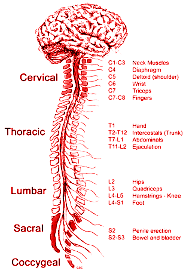
The human spinal cord is typically 40 cm long and 2 cm wide in adults, extending from the medulla oblongata to the lumbar region of the vertebral column. It is divided into five main regions:
Cervical cord – Located in the neck region.
Thoracic cord – Found in the upper and mid-back.
Lumbar cord – Situated in the lower back.
Sacral cord – At the base of the spine.
Coccygeal – The tail end of the spinal cord.
Each region contains spinal nerves that connect to different parts of the body. There are 31 pairs of spinal nerves, classified as follows:
8 cervical nerves (C1-C8)
12 thoracic nerves (T1-T12)
5 lumbar nerves (L1-L5)
5 sacral nerves (S1-S5)
1 coccygeal nerve (Co1)
These spinal nerves transmit signals between the brain and body, controlling reflexes, muscle movements, and sensory functions.
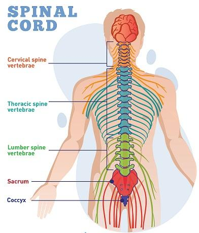
Spinal Cord Diagram – Labelled and Easy
A labelled spinal cord diagram helps in better visualising its structure. Below is an easy-to-understand spinal cord diagram that shows its different parts.

Key Points:
The spinal cord has different segments, each with specific nerve connections.
It is housed within the vertebral column, which protects it from injuries.
Grey and white matter play distinct roles in sensory and motor functions.
Structure of the Spinal Cord
The spinal cord runs through a hollow cavity inside the vertebral column, protected by three layers of meninges:
Dura mater – The outermost layer.
Arachnoid mater – The middle protective layer.
Pia mater – The innermost layer, closely attached to the spinal cord.
Cross-section of the Spinal Cord
A cross-section of the spinal cord reveals grey and white matter:
Grey matter – Located in the centre, shaped like a butterfly. It contains nerve cells and the central canal filled with cerebrospinal fluid (CSF).
White matter – Surrounds the grey matter, consisting of axons that transmit nerve signals.
The spinal cord structure and function are crucial for relaying sensory (afferent) signals to the brain and motor (efferent) signals from the brain to the body.

Spinal Cord Function
The spinal cord serves as a vital link between the brain and body, performing multiple functions:
Relays messages between the brain and the body.
Controls reflex actions without brain involvement.
Provides structural support to maintain body posture.
Coordinates movement and muscle control.
Regulates autonomic functions, such as heart rate and digestion.
Facilitates pain and temperature sensation transmission.
The spinal cord function is crucial in coordinating reflexes and ensuring smooth bodily movements.
Spinal Cord Injuries
Damage to the spinal cord can cause severe health issues due to the loss of communication between the brain and body. Injuries can be classified as:
1. Complete Injury
Total loss of motor and sensory function below the injury site.
Results in paralysis of affected body parts.
2. Incomplete Injury
Partial loss of function, with some movement or sensation still present.
Types of Paralysis Due to Spinal Cord Injury
Tetraplegia (Quadriplegia) – Affects all four limbs and torso.
Paraplegia – Affects the lower limbs and lower body.
Symptoms include:
Loss of voluntary movements
Numbness or loss of sensation
Reflex abnormalities
Breathing difficulties in severe cases
Early diagnosis and treatment can help in managing spinal cord injuries effectively.
Spinal Cord Nerves & Types
The 31 pairs of spinal nerves are divided into different categories based on their location:
Cervical Nerves (C1-C8) – Control neck, arms, and diaphragm movements.
Thoracic Nerves (T1-T12) – Control chest and upper abdominal muscles.
Lumbar Nerves (L1-L5) – Control lower back, hips, and leg functions.
Sacral Nerves (S1-S5) – Control pelvic organs, lower limbs, and feet.
Coccygeal Nerve (Co1) – Controls the tailbone region.
These spinal cord nerves play a significant role in maintaining motor and sensory functions throughout the body.
Unique Information
1. Role of the Spinal Cord in Reflex Actions: The spinal cord is responsible for reflexes, which are automatic responses to stimuli. The reflex arc involves sensory neurons, interneurons, and motor neurons to execute quick responses.
2. How the Spinal Cord Adapts to Injuries: In some cases, the spinal cord can retrain the brain to use alternative pathways for nerve signal transmission, helping people recover partial movements after injuries.
3. The Spinal Cord and Neuroplasticity: Neuroplasticity enables the spinal cord to form new neural connections even after minor injuries, improving recovery chances.
Related Links:


FAQs on Spinal Cord: Structure, Functions, and Detailed Diagram
1. What are the main functions of the spinal cord in our daily activities?
The spinal cord acts as the body's central information highway. Its main functions are:
- Signal Transmission: It carries nerve signals from the brain to the rest of the body to control movement, and from the body back to the brain to register sensations like touch and heat.
- Reflex Control: It manages reflex actions, which are automatic, rapid responses to stimuli (like pulling your hand away from a hot object) that happen without involving the brain first.
2. How is the spinal cord protected inside our body?
The spinal cord is very delicate and has multiple layers of protection. It is shielded by the vertebral column (backbone), a series of bones called vertebrae. Additionally, it is covered by three protective membranes known as meninges and cushioned by a special fluid called cerebrospinal fluid, which absorbs shocks.
3. What are the key parts to label in a diagram of the spinal cord's cross-section?
For a Class 10 level diagram, the most important parts to label in a cross-section of the spinal cord are the central H-shaped grey matter, the surrounding white matter, the central canal (a small channel in the middle), and the emerging spinal nerves with their dorsal and ventral roots.
4. What is the main difference between the grey matter and white matter in the spinal cord?
The main difference lies in their composition and function. Grey matter, which forms the H-shape in the center, contains neuron cell bodies and is where information processing happens. White matter surrounds the grey matter and is made of nerve fibres (axons) with a fatty coating, which allows it to transmit signals quickly up and down the spinal cord.
5. How does the spinal cord manage a reflex action so quickly, even before the brain is aware?
The spinal cord manages this through a direct pathway called the reflex arc. When a sensory receptor detects a danger (like extreme heat), the signal travels to the spinal cord. Here, an interneuron immediately passes the signal to a motor neuron, which triggers a muscle response. This shortcut allows for an instant reaction, and the signal is sent to the brain only after the action has already occurred.
6. Why can an injury to the spinal cord cause someone to lose feeling and movement in their legs?
An injury can cause paralysis because the spinal cord is the only path for communication between the brain and the lower body. If the cord is damaged, the nerve signals from the brain that command the legs to move cannot get through. Similarly, sensory signals from the legs, like touch or pain, cannot travel up to the brain to be processed, leading to a loss of feeling.


