


Placenta Functions in Human Pregnancy for NEET Biology Students
Placenta is a crucial biological structure in mammals, acting as the lifeline between mother and developing fetus. For NEET aspirants, understanding the placenta is essential as it connects key principles of human physiology, reproduction, and embryology. This topic appears frequently in NEET and forms the foundation for grasping fetal development and maternal-fetal interactions in Biology.
What is Placenta? (Placenta Meaning and Definition)
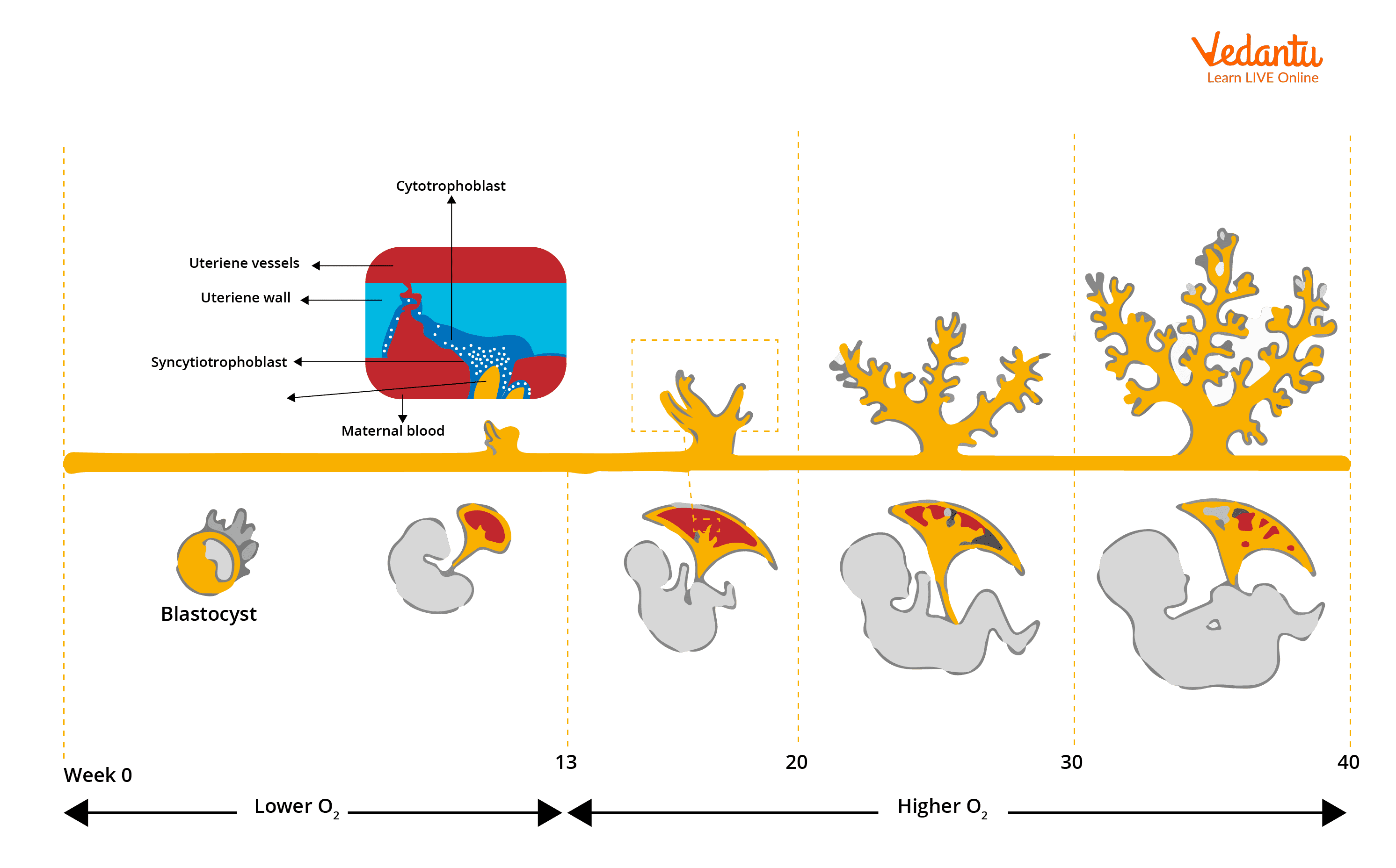
The placenta is a temporary organ that develops during pregnancy in mammals. It forms at the interface of maternal uterine tissue and the growing fetus, serving as a highly specialized structure for exchanging nutrients, gases, and wastes between mother and fetus. The placenta ensures that the developing fetus receives oxygen and nutrients while simultaneously removing waste products. In humans, it forms shortly after implantation and remains functional until birth, after which it is expelled as "afterbirth."
Core Ideas and Fundamentals of Placenta
Structure of Human Placenta
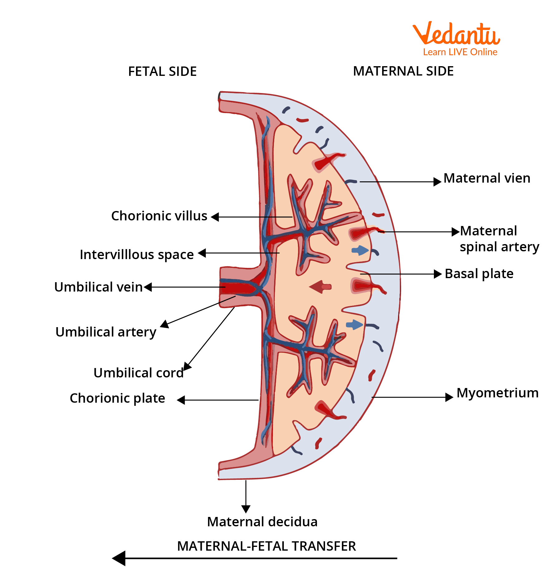
The human placenta is a disc-shaped organ, rich in blood supply, weighing about 500 grams at full term. It consists of fetal and maternal components, intricately interwoven but separated by a thin barrier. The fetal side forms from chorionic villi, while the maternal part derives from endometrial tissue. This unique structure allows selective transfer of substances and acts as a protective filter.
Functions of the Placenta
- Supplies oxygen and nutrients from the mother's blood to the fetus.
- Removes carbon dioxide and metabolic waste from fetal blood.
- Serves as an endocrine organ, producing hormones like hCG, progesterone, and estrogen, which regulate pregnancy.
- Provides immune protection to the fetus by blocking some harmful substances and pathogens.
- Acts as a selective barrier, allowing or preventing passage of certain materials.
Formation and Development of the Placenta
Placental development begins soon after fertilized egg implantation. Chorionic villi sprout from the trophoblast and invade the maternal endometrium, establishing contact with maternal blood vessels. This connection matures over weeks, creating the functional placenta.
Important Sub-Concepts Related to Placenta
Types of Placenta
On the basis of tissue separation between maternal and fetal blood, placentas are categorized into types like epitheliochorial, endotheliochorial, and haemochorial. Humans possess a haemochorial placenta, allowing direct contact between fetal chorionic villi and maternal blood, ensuring efficient exchange.
Placental Hormones
- hCG (Human Chorionic Gonadotropin): Maintains corpus luteum function in early pregnancy.
- Progesterone: Maintains uterine lining to support pregnancy.
- Estrogen: Promotes uterine blood flow and fetal organ development.
- hPL (Human Placental Lactogen): Prepares maternal body for lactation and modulates maternal metabolism.
Key Principles and Relationships Related to Placenta
Understanding how exchange occurs across the placental barrier is vital. Simple diffusion, facilitated diffusion, active transport, and pinocytosis are the main mechanisms.
| Mode of Transport | Substances Transferred | Example |
|---|---|---|
| Simple Diffusion | Gases | Oxygen, Carbon dioxide |
| Facilitated Diffusion | Nutrients | Glucose |
| Active Transport | Ions | Calcium, Iron |
| Pinocytosis | Large Molecules | Immunoglobulin G (IgG) |
This table summarizes how vital substances are exchanged through different processes across the placenta, which is a frequent NEET concept question area.
Features and Limitations of Human Placenta
Key Features
- Highly vascular, maximizing efficiency of exchange.
- Produces several crucial hormones for pregnancy maintenance.
- Acts as both a physical and biochemical barrier.
Limitations and Challenges
- Cannot block all toxins, drugs, or infectious agents (teratogens like alcohol or rubella can cross).
- Some maternal antibodies may attack fetal tissues in case of Rh incompatibility.
Why is Placenta Important for NEET?
Placenta is a high-yield topic in NEET Biology, commonly appearing in Reproduction in Humans, Human Physiology, and Embryology. Understanding it helps clarify fundamental processes like fetal nutrition, gas exchange, and hormonal regulation. Questions often test conceptual clarity, clinical implications (like placental insufficiency), and even comparative types across mammals. It also connects with related NEET topics such as fertilization, implantation, and hormonal cycles. Mastery over this topic supports both direct MCQ solving and applied reasoning in assertion-reason type questions.
How to Study Placenta Effectively for NEET
- Start by visualizing diagrams of placenta structure and development. Regularly label and revise them.
- Focus on understanding physiological functions, not just rote learning names.
- Memorize key hormones, their sources, and roles. Make a comparative table if possible.
- Practice MCQs that ask about structure, function, and clinical significance of placenta.
- Connect placental details with broader concepts in reproduction and fetal development for better retention.
- After completing theoretical study, attempt assertion-reason and diagram-based questions.
- Revise by summarizing mechanisms of substance exchange and hormone actions.
Common Mistakes Students Make in Placenta
- Confusing maternal and fetal parts or failing to identify which tissues are of maternal origin versus fetal origin.
- Incorrectly remembering the types and classification of placentas among different mammals.
- Mixing up functions of placental hormones, especially hCG, hPL, progesterone, and estrogen.
- Ignoring the details of barrier functions and mistakenly assuming all substances can cross easily.
- Overlooking clinical significance, such as what happens in Rh incompatibility or placental disorders.
Quick Revision Points on Placenta
- Placenta connects fetal and maternal blood for nutrient, gas, and waste exchange.
- Human placenta is haemochorial type.
- Produces vital hormones for pregnancy maintenance (e.g., hCG, progesterone).
- Acts as a selective barrier, provides immune protection, but some harmful substances can cross.
- Main transport mechanisms: diffusion, facilitated diffusion, active transport, pinocytosis.
- Study structure, function, development, and hormonal aspects for NEET.
- Revise diagrams and key functions before the exam.


FAQs on Placenta in NEET Biology: Structure and Functions
1. What is placenta and what is its function in pregnancy for NEET?
The placenta is a temporary organ that develops during pregnancy and provides nutrients, oxygen, and waste removal for the developing fetus. Key NEET points include:
- Exchange of nutrients and gases: Transfers oxygen and nutrients from the mother’s blood to the fetus.
- Waste elimination: Removes carbon dioxide and fetal wastes into the mother’s bloodstream.
- Hormone production: Secretes hormones such as hCG, estrogen, and progesterone to maintain pregnancy.
- Protection: Acts as a barrier to some harmful substances, though not all toxins or pathogens.
Understanding placenta structure and function is important for NEET biology exams.
2. What are the main functions of the placenta according to the NEET syllabus?
The main functions of the placenta are vital for fetal development in pregnancy. For NEET, focus on:
- Nutrient supply (glucose, amino acids, fatty acids, vitamins)
- Respiratory exchange (oxygen delivery, carbon dioxide removal)
- Excretion of waste products (urea, creatinine)
- Endocrine role (producing hCG, estrogen, progesterone)
- Barrier function against some toxins and microorganisms
Review all these for NEET biology as they're frequently asked in exams.
3. What is the structure of the placenta in humans for NEET?
The human placenta has a discoid and hemochorial structure, crucial for NEET exams:
- Discoid shape: Flattened, disc-like organ attached to the uterine wall.
- Hemochorial type: Maternal blood comes in direct contact with the chorionic villi.
- Two main components: Maternal part (decidua basalis) and fetal part (chorion frondosum).
- Chorionic villi: Increase the surface area for exchange of materials.
Remember details of placenta structure for NEET-based MCQs.
4. What hormones are secreted by the placenta during pregnancy?
The placenta secretes several hormones essential for maintaining pregnancy, often asked in NEET:
- hCG (human chorionic gonadotropin): Supports the corpus luteum.
- Progesterone: Maintains the uterine lining.
- Estrogens: Promote uterine growth and mammary gland development.
- hPL (human placental lactogen): Stimulates breast development and regulates fetal metabolism.
These hormones are noteworthy for the NEET examination, particularly in the physiology of pregnancy.
5. What is the role of placenta in exchange of gases?
The placenta facilitates the exchange of respiratory gases between mother and fetus, an important NEET concept:
- Oxygen diffuses from maternal to fetal blood through the placenta.
- Carbon dioxide returns from the fetal blood to the maternal circulation for removal.
- This gas exchange ensures proper fetal development and is achieved via placental villi.
Placental gas exchange is a key topic for NEET biology questions on fetal physiology.
6. How does the placenta protect the fetus from harmful substances for NEET?
The placenta acts as a partial selective barrier, protecting the fetus from some harmful substances. Key NEET points include:
- Blocks large molecules, certain bacteria, and some toxins.
- Allows passage of nutrients, gases, and some antibodies (IgG).
- Does not protect against all viruses, alcohol, drugs, and some chemicals.
For NEET, know the limits of placental barrier function and its role in fetal health.
7. Why is placenta called a temporary endocrine gland in NEET biology?
The placenta is called a temporary endocrine gland because it produces hormones only during pregnancy:
- Secretes hCG, estrogen, progesterone until birth.
- Regulates and maintains pregnancy and fetal development.
- Dissolves after delivery and ceases hormone production.
This concept is frequently tested in NEET endocrine system questions.
8. What are the differences between maternal and fetal sides of the placenta?
The placenta has distinct maternal and fetal portions, which is a common NEET query:
- Maternal side: Called decidua basalis, attached to the uterus, rich in maternal blood.
- Fetal side: Formed by chorion frondosum, contains chorionic villi and umbilical cord attachment.
- Both sides work together to enable nutrient and gas exchange.
Identifying these differences is important for NEET exam MCQs about placental anatomy.
9. What will happen if the placenta does not function properly during pregnancy?
Abnormal placenta function can lead to serious complications for both mother and fetus, a NEET-relevant topic:
- Fetal growth restriction: Inadequate nutrients and oxygen supply.
- Pre-eclampsia: High blood pressure and proteinuria in the mother.
- Preterm birth or miscarriage: Due to insufficient hormonal support.
Understanding placenta-related disorders is essential for NEET syllabi regarding pregnancy health.
10. What is the importance of placenta for fetal development?
The placenta is essential for supporting, protecting, and nourishing the developing fetus throughout pregnancy. Major points for NEET students to remember are:
- It supplies oxygen and nutrients from mother to baby.
- Removes carbon dioxide and waste from fetal blood.
- Produces key hormones needed during pregnancy.
- Acts as a defense system for the fetus against some infections and toxins.
Knowing the importance of the placenta is crucial for NEET exam preparation in human reproduction.
11. Define placenta in one line for NEET.
The placenta is a temporary, disc-shaped organ that forms during pregnancy, connecting the developing fetus to the maternal uterus for exchange of nutrients, gases, and wastes.
This concise definition is suitable for NEET one-mark questions.
12. What are the types of placenta based on structure?
Based on structure, placenta types include:
- Discoidal placenta: Found in humans and rodents.
- Zonary placenta: Present in dogs and cats.
- Cotyledonary placenta: Found in ruminants like cows and sheep.
- Diffuse placenta: Seen in horses and pigs.
NEET often focuses on discoidal, hemochorial placenta in humans.