Functions of Human Ear Parts with Diagram for NEET & CBSE Students
The human ear is a vital sensory organ in our body, responsible not only for detecting and interpreting sound but also for maintaining balance and spatial orientation. Understanding its structure, labelled parts, and key functions is essential for students in school biology, as well as for parents and teachers helping guide these concepts. The ear’s anatomy can be divided into three main sections: the outer ear, middle ear, and inner ear. Each section plays a unique role, working together to help us hear and stay balanced in our daily activities.
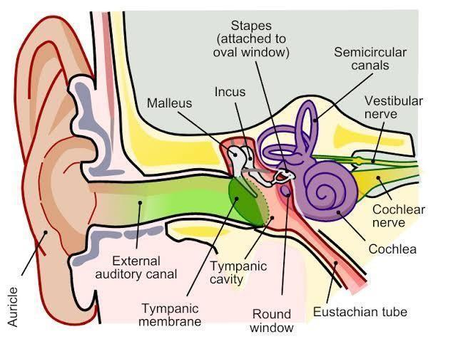
Structure of the Human Ear – Overview and Labelling
The ear is structured in three main regions:
- Outer Ear: The most visible part, including the pinna (auricle) and the external auditory canal. The pinna collects sound waves and funnels them into the ear canal.
- Middle Ear: Begins at the tympanic membrane (eardrum) and contains three small interconnected bones called ossicles (malleus, incus, stapes). These amplify and transmit vibrations to the inner ear. The Eustachian tube connects the middle ear to the throat, equalizing air pressure.
- Inner Ear: Comprises the cochlea (responsible for hearing) and the vestibular apparatus (semicircular canals and vestibule) that control balance.

Detailed Breakdown of Human Ear Parts
Let’s understand the function and significance of each part:
- Pinna (Auricle): Outer, flap-like structure that collects sound and helps determine the direction of incoming noise.
- External Auditory Canal: Channel for sound waves; contains protective glands and fine hairs.
- Tympanic Membrane (Eardrum): A thin barrier, vibrating on impact with sound waves and initiating the hearing process.
- Ossicles (Malleus, Incus, Stapes): The smallest bones in our body. They act as transmitters, amplifying sound vibrations toward the inner ear.
- Eustachian Tube: Maintains equal air pressure on both sides of the eardrum, enabling accurate vibration transmission. Swallowing opens this tube, balancing pressure.
- Cochlea: A spiral, fluid-filled organ where vibrations are converted into nerve impulses by hair cells. Essential for hearing various pitches.
- Semicircular Canals: Three fluid-filled loops, each positioned at right angles, crucial for detecting head rotation and balance.
- Vestibule: The central area of the inner ear that senses linear movements and gravity.
- Auditory Nerve: Carries processed sound and balance information from the inner ear to the brain.
Step-by-Step Process: How Does the Ear Function?
1. Sound waves enter the pinna and move through the auditory canal.
2. They reach the tympanic membrane, making it vibrate.
3. The ossicles amplify and convey these vibrations to the inner ear.
4. In the cochlea, mechanical vibrations are turned into electrical impulses.
5. The auditory nerve transmits these signals to the brain for sound recognition.
6. The semicircular canals and vestibule send information about body movement and position to maintain balance.
Key Scientific Definitions and Importance
- Tympanic Membrane: The boundary between the outer and middle ear, critical for sound transmission.
- Ossicles: Malleus (hammer), incus (anvil), and stapes (stirrup) — amplifier bones central to hearing.
- Cochlea: The actual ‘organ of hearing’, spiral-shaped to fit more sensory cells within a small area.
- Eustachian Tube: Pressure regulator, ensuring undistorted hearing.
- Semicircular Canals and Vestibule: Work as our internal gyroscope, preventing dizziness and imbalance.
| Region | Main Components | Function |
|---|---|---|
| Outer Ear | Pinna, Auditory Canal | Collects and channels sound |
| Middle Ear | Eardrum, Ossicles, Eustachian Tube | Amplifies and transmits sound vibrations |
| Inner Ear | Cochlea, Semicircular Canals, Vestibule, Auditory Nerve | Converts sound to nerve signals, controls balance |
Exam Tip: Mnemonic for Ear Bones
A quick way to remember the order of ossicles from the eardrum inward: My Inner Spaces
I – Incus
S – Stapes
Practice Questions – Test Your Knowledge
- Draw and label a diagram of the human ear. Briefly describe each part’s main function.
- Explain how the cochlea and semicircular canals differ in their function and structure.
- How does the Eustachian tube assist in the process of hearing?
- List the sequence of events as sound travels through the ear, starting from the pinna.
Next Steps and Learning Resources
- Read more on human ear structure and functions to understand related topics.
- Deepen your learning about the anatomy of the ear and inner ear in further chapters.
- For targeted practice, try labelling blank diagrams and answering past-year questions on ear anatomy.
Understanding the labelled structure of the human ear builds a clear foundation for further study in biology, medicine, and everyday awareness of hearing and balance health. Use diagrams, practice questions, and Vedantu’s linked resources to reinforce your knowledge and confidence.


FAQs on Human Ear Labelled Diagram: Parts, Structure, and Functions
1. What are the main parts of the human ear in a labelled diagram?
The main parts of the human ear shown in a labelled diagram are:
- Outer ear: Pinna and auditory canal
- Middle ear: Tympanic membrane (ear drum), ossicles (malleus, incus, stapes), and Eustachian tube
- Inner ear: Cochlea, vestibular apparatus (semicircular canals and vestibule), and auditory nerve
Each part is essential for hearing and balance.
2. How do you draw a labelled diagram of the human ear?
To draw a labelled diagram of the human ear:
- Begin with an outline of the ear showing three regions: outer, middle, and inner.
- Mark and label: pinna, auditory canal, tympanic membrane, ossicles (malleus, incus, stapes), Eustachian tube, cochlea, semicircular canals, vestibule, and auditory nerve.
- Use straight lines for labels and neatly write each part's name.
Practicing NCERT-based diagrams helps in exams.
3. What are the functions of the main parts of the human ear?
The human ear performs key functions through its distinct parts:
- Outer ear: Collects and funnels sound waves
- Middle ear: Amplifies vibrations and equalizes pressure
- Inner ear: Converts vibrations to nerve impulses (hearing) and maintains balance
Each part has a specific physiological role critical for auditory perception and balance.
4. Why is the cochlea called the 'organ of hearing'?
The cochlea is called the 'organ of hearing' because it contains the organ of Corti, which:
- Has hair cells that detect sound vibrations
- Converts these vibrations into nerve impulses
- Sends the impulses via the auditory nerve to the brain for sound perception
The cochlea's unique spiral structure makes it vital for hearing.
5. What is the function of the semicircular canals in the ear?
The semicircular canals are part of the vestibular apparatus in the inner ear. Their main function is to:
- Detect changes in head position and movement
- Maintain body balance and spatial orientation
- Send signals to the brain about equilibrium
They are essential for maintaining balance and coordination during movement.
6. How does the Eustachian tube help in ear function?
The Eustachian tube connects the middle ear to the pharynx. It:
- Equalizes air pressure on both sides of the tympanic membrane
- Prevents damage to the ear drum
- Helps maintain normal hearing
This tube opens during swallowing or yawning to balance ear pressure.
7. What is the path of sound through the human ear?
Sound travels through the ear in a specific path:
1. Sound waves enter the outer ear (pinna, auditory canal).
2. Strike the tympanic membrane, causing it to vibrate.
3. Vibrations pass through the ossicles (malleus, incus, stapes) in the middle ear.
4. Vibrations reach the cochlea in the inner ear.
5. Hair cells in the cochlea convert vibrations into nerve impulses.
6. Impulses travel via the auditory nerve to the brain.
This pathway is essential for hearing and interpreting sounds.
8. How does the ear help maintain balance?
The human ear maintains balance through the vestibular apparatus in the inner ear.
- The semicircular canals detect rotational movements.
- The vestibule senses changes in head position and acceleration.
- Sensory cells send signals to the brain to help control posture and equilibrium.
This system ensures stability during movement and changes in body position.
9. Why is labelling important in the diagram of the human ear?
Labelling in the diagram of the human ear is important because:
- It helps identify and memorize key structures for exams
- Clarifies the function of each part (e.g., cochlea for hearing, semicircular canals for balance)
- Enables quick revision and boosts accuracy in diagram-based questions
Accurate labelling follows the NCERT and NEET exam pattern.
10. What type of questions can be asked from the human ear diagram in NEET or boards?
Typical exam questions from the human ear diagram include:
- Label the given human ear diagram and write functions of each part
- Identify outer, middle, and inner ear regions
- Explain the process of hearing using a flowchart or diagram
- Describe differences between cochlea and semicircular canals
Diagram labelling and functional explanation are regularly asked in NEET, CBSE, and ICSE exams.
11. What are ossicles and what is their function?
Ossicles are three small bones in the middle ear: malleus, incus, and stapes.
- They amplify and transfer sound vibrations from the tympanic membrane to the inner ear (cochlea).
- Proper ossicle function is critical for efficient hearing.
The stapes is the smallest bone in the human body.
12. What is the difference between the cochlea and vestibule?
The cochlea and vestibule are both part of the inner ear, but have different functions:
- Cochlea: Spiral-shaped, responsible for converting sound vibrations into electrical signals (hearing).
- Vestibule: Central part of the bony labyrinth, involved in balancing and detecting linear movements.
Both structures are essential for hearing and equilibrium but serve distinct roles.


