

How to Draw and Label a Chromosome Diagram for Exams
The concept of chromosome diagram is essential in biology and helps explain real-world biological processes and exam-level questions effectively. A labeled chromosome diagram clearly shows the structure, anatomy, and major parts of a chromosome, making it easier for students to understand genetic inheritance, mutation, and cell division. This topic is frequently tested in board exams and is vital for Class 10, 11, and 12 Biology students.
Understanding Chromosome Diagram
Chromosome diagram refers to a detailed, labeled drawing that illustrates the structure of a chromosome inside the cell nucleus. This concept is important in areas like chromosome structure & function, genetic inheritance, and cell division processes. Chromosome diagrams help visualize key features like the centromere, chromatids, telomeres, and arms, all of which play vital roles during mitosis and meiosis.
Key Parts of a Chromosome Diagram
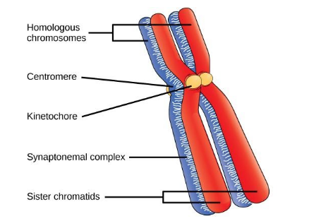
A chromosome diagram usually shows these major parts:
- Chromatids: Two identical halves of a chromosome, visible during cell division.
- Centromere: The constricted region joining the two chromatids and dividing the chromosome into two arms.
- Arms (p and q): The shorter arm is called 'p', and the longer arm is 'q'.
- Telomere: The terminal end portions of the chromosome protecting it from fusion with other chromosomes.
- Kinetochore: A disc-shaped protein on the centromere where spindle fibers attach during cell division.
- Satellite: Sometimes found at the end of a chromosome, connected by a thin filament.
Types of Chromosome Based on Centromere Position
The position of the centromere gives chromosomes their characteristic shapes. Here’s a helpful table to understand chromosome types better:
Types of Chromosome Diagram
| Type | Centromere Position | Shape |
|---|---|---|
| Metacentric | Centromere in the middle | V-shaped, arms equal length |
| Submetacentric | Centromere slightly off center | J or L-shaped, arms unequal |
| Acrocentric | Centromere close to one end | Rod-like, one very short arm |
| Telocentric | Centromere at the terminal end | I-shaped, arms on one side only |
How to Draw & Label a Chromosome Diagram
Drawing a chromosome diagram correctly is crucial for exam marks. Here’s a simple step-by-step guide:
- Draw an X-shape (for replicated chromosome) or a rod for single chromatid.
- Mark the constriction (centromere) joining the two arms.
- Label the 'p' (short) and 'q' (long) arms on either side of the centromere.
- Point out telomeres at both ends.
- Label chromatids and, if relevant, mark the position of alleles or genes.
- Add the kinetochore on the centromere, if needed for class 11/12.
Chromosome vs. Chromatin vs. DNA Diagram
Students often get confused between chromosome, chromatin, and DNA in diagrams:
| Component | Appearance | When Visible |
|---|---|---|
| Chromosome | Condensed X-shaped | During cell division |
| Chromatin | Loose, thread-like | In interphase (resting cell) |
| DNA | Double helix, molecular view | At all times (invisible with light microscope) |
Class-wise Chromosome Diagrams for Exams
Board exams for classes 10, 11, and 12 frequently require students to draw and label a chromosome diagram. Class 10 students may be asked for simpler labeling (centromere, arms), while class 11/12 might require alleles, satellite, and kinetochore. Practicing different diagrams improves accuracy and exam marks.
Common Mistakes to Avoid
- Confusing chromosome diagram with chromatin or DNA structures.
- Missing key labels such as the centromere or mislabeling arms.
- Not showing both chromatids in metaphase diagrams.
Real-World Applications
The concept of chromosome diagram is used in genetics, medicine, and biotechnology. Clear chromosome diagrams help in understanding genetic diseases (like Down’s syndrome), analyzing karyotypes, and during advanced genetics research. Vedantu helps students relate these diagrams to real class experiments and practical biology.
Practice Questions
- What are the main parts labeled in a standard chromosome diagram?
- Explain the differences between metacentric and acrocentric chromosomes with diagrams.
- How is the position of the centromere shown in a chromosome diagram?
- Draw and label a chromosome diagram showing genes and alleles.
Explore Related Concepts
- Chromosome
- Difference Between Gene and Chromosome
- Cell Structure and Function
- Nucleus
- DNA Structure
- Chromatin
- Cell Cycle
- Differences Between Mitosis and Meiosis
In this article, we explored chromosome diagram, its key processes, real-life significance, and how to solve questions based on it. To learn more and build confidence, keep practicing with Vedantu’s structured biology resources and diagrams.


FAQs on Chromosome Diagram: Parts, Structure, and Stepwise Drawing
1. What is a chromosome diagram?
A chromosome diagram is a labeled, visual representation that illustrates the structure, parts (such as the centromere, chromatids, and telomeres), and the biological function of a chromosome within a cell. It helps students understand chromosome anatomy, facilitating easier exam preparation and concept retention.
2. How do you label a chromosome diagram?
To label a chromosome diagram, follow these steps:
1. Identify and mark the centromere, which joins the chromatids.
2. Label each chromatid as the identical arms of the chromosome.
3. Mark the short 'p' arm and the long 'q' arm.
4. Indicate the telomeres at the ends of chromatids.
5. Optionally, include structures like satellites or secondary constrictions based on syllabus requirements.
Clear labels with pointers or arrows help demonstrate the chromosome’s parts effectively.
3. What are the 4 main parts of a chromosome?
The four key parts of a chromosome are:
• The centromere — the central region joining two chromatids.
• The two chromatids — each containing a DNA molecule.
• The telomeres — protective end caps of the chromosome.
• The arms — the shorter p arm and the longer q arm.
These parts collectively form the overall structure vital for genetic stability and cell division.
4. What is the function of a centromere in a chromosome diagram?
The centromere acts as the attachment point for sister chromatids, holding them together until they separate during anaphase of cell division. It also facilitates the assembly of the kinetochore, which binds spindle fibers essential for accurate chromosome segregation, ensuring proper distribution of genetic material.
5. How do chromosomes differ from chromatin and DNA in diagrams?
In diagrams, chromosomes appear as condensed, X-shaped structures visible during cell division. Chromatin is a loosely packed complex of DNA and proteins present in the nucleus during interphase. DNA is the molecular blueprint tightly coiled into chromatin and further condensed to form chromosomes, showing different organizational levels in cellular diagrams.
6. What are the 4 types of chromosomes based on centromere position?
Chromosomes are classified by their centromere position into:
• Metacentric — centromere in the middle, arms of equal length.
• Submetacentric — centromere slightly off-center, unequal arms.
• Acrocentric — centromere near one end, resulting in a very short and a very long arm.
• Telocentric — centromere at the very end, giving an I-shaped chromosome.
This classification aids in identifying chromosome shape during cytogenetic analysis and exams.
7. Why are some chromosome diagrams shown as X-shaped?
Chromosome diagrams show the characteristic X-shape during the metaphase stage of mitosis and meiosis, when chromosomes are fully condensed and consist of two sister chromatids joined at the centromere. This shape represents the duplicated status of chromosomes prior to their separation into daughter cells.
8. Why do students confuse labeling between chromatids and centromeres?
Students often confuse chromatids and centromeres because diagrams frequently depict closely positioned parts and similar terminology. The chromatids are the individual arms or strands holding identical DNA copies, whereas the centromere is the central constriction joining chromatids. Clear stepwise labeling and diagram practice help overcome this confusion.
9. Why are accurate diagrams important for CBSE and NEET exams?
Accurate, well-labeled diagrams are critical in CBSE and NEET exams as they demonstrate a clear understanding of important biological structures. They help secure valuable marks, improve clarity in answers, and ensure compliance with prescribed syllabus guidelines. Precise diagramming also aids in grasping complex concepts and supports effective revision.
10. How do alleles appear visually on a chromosome diagram?
In chromosome diagrams, alleles are represented as specific loci or positions on the chromatids where genes reside. Though not visually distinct at basic diagram levels, advanced diagrams may show alleles as paired markers or labels on homologous chromosomes, illustrating genetic variation and the inheritance of traits.
11. Why is the chromosome diagram different for mitosis and meiosis?
Chromosome diagrams differ between mitosis and meiosis because of the stages and structural changes they depict. In mitosis, chromosomes appear as duplicated chromatids separating equally, while meiosis involves homologous chromosome pairing, crossing over, and reduction division. Diagrams highlight these unique features to explain different cell division outcomes.


