


Complete Guide to Meiosis Cell Division, Phases of Meiosis, Stages and Genetic Significance
Meiosis is a special type of cell division that reduces the chromosome number by half and produces haploid cells from a diploid parent cell. In humans and many other sexually reproducing organisms, this process is essential for the formation of gametes such as sperm and eggs.
Unlike mitosis, which produces genetically identical daughter cells for growth and repair, meiosis is meant for sexual reproduction and genetic variation.
Meiosis is a reductional division in which one diploid cell gives rise to four haploid cells. It is called reductional because the chromosome number is reduced from diploid (2n) to haploid (n).
In sexually reproducing organisms, meiosis ensures that chromosome number remains constant from one generation to the next. If meiosis did not occur, fertilization would continuously double the chromosome number in every generation.
Main Purpose of Meiosis
Formation of gametes
Reduction of chromosome number
Creation of genetic variation
Maintenance of chromosome number across generations
In humans, meiosis occurs in the reproductive organs and produces:
Sperm cells in males
Egg cells in females
Meiosis Cell Division: Why It Is Unique?
Meiosis cell division is unique because the cell must perform two major tasks:
Separate homologous chromosomes
Separate sister chromatids
This is different from mitosis, where only sister chromatids are separated.
The two-step nature of meiosis helps achieve this:
Meiosis I separates homologous chromosomes
Meiosis II separates sister chromatids
As a result, one starting cell produces four daughter cells instead of two. These daughter cells are haploid and genetically different from each other.
Overview of Meiosis Division
The entire meiosis division can be divided into:
Interphase
Meiosis I
Meiosis II
Each meiotic division contains four stages:
Prophase
Metaphase
Anaphase
Telophase
Interphase Before Meiosis
Before entering meiosis I, the cell undergoes interphase. This stage is essential because the cell prepares itself for division.
Interphase includes:
G₁ phase – cell grows
S phase – DNA replicates
G₂ phase – cell prepares for division
This means that although meiosis ultimately produces haploid cells, DNA replication happens only once, before meiosis I begins. There is no DNA replication between meiosis I and meiosis II.
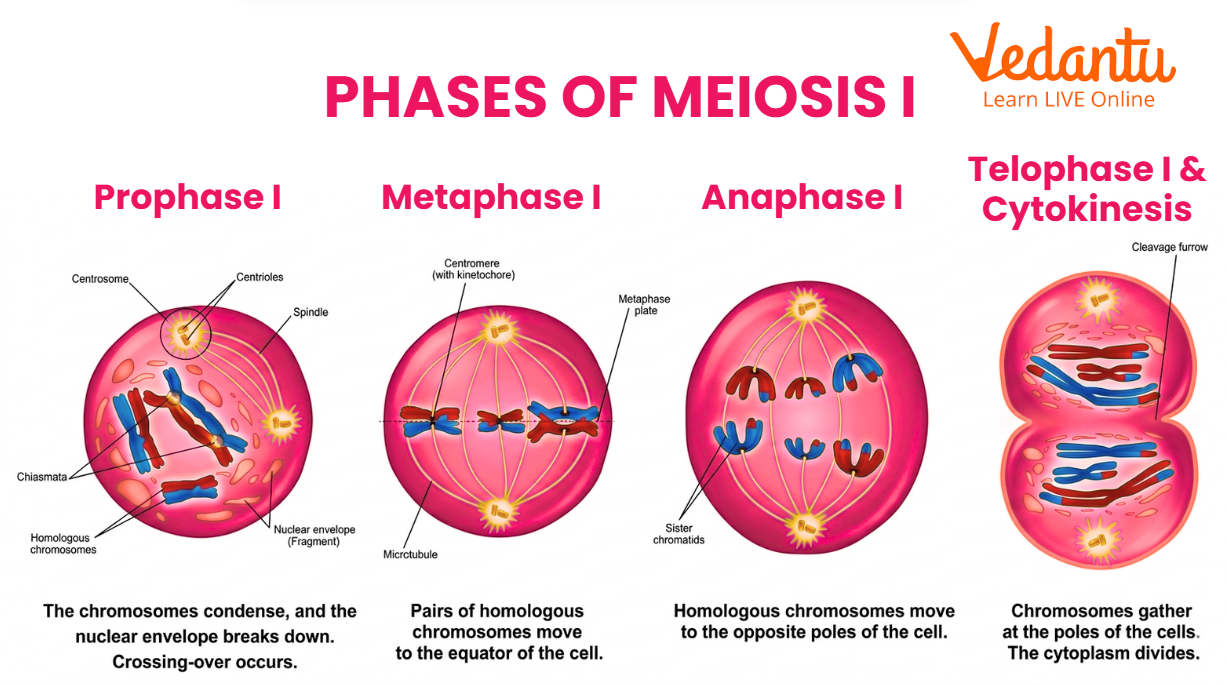
Meiosis I
Meiosis I is the first meiotic division and is called the reductional division because it reduces chromosome number by separating homologous chromosomes.
This is the more complex part of meiosis because homologous chromosomes must:
Pair with each other
Exchange segments
Separate into different cells
The four stages of meiosis I are described below.
Prophase I
Prophase I is the longest and most important stage of meiosis. It is here that meiosis becomes clearly different from mitosis.
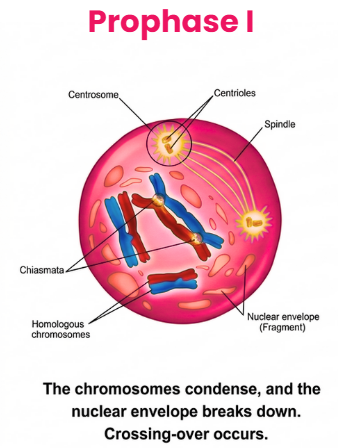
Major Events of Prophase I
1. Chromosome condensation
Chromosomes begin to condense and become visible under the microscope.
2. Pairing of homologous chromosomes
Each chromosome pairs with its corresponding homologue. One member of each pair comes from the mother and the other from the father. This pairing is called synapsis.
3. Formation of synaptonemal complex
A protein structure called the synaptonemal complex holds homologous chromosomes together closely during pairing and crossing over.
4. Crossing over
Homologous chromosomes exchange corresponding segments of DNA. This process is called crossing over. It produces new combinations of alleles and increases genetic variation.
5. Formation of chiasmata
The visible points where crossing over has occurred are called chiasmata. These maintain the association between homologous chromosomes even after the synaptonemal complex disappears.
Why Prophase I is important?
Prophase I is the most significant meiotic stage because:
homologous chromosomes pair only in meiosis
crossing over occurs only in meiosis
genetic recombination begins here
These features make meiosis essential for evolution and variation.
Metaphase I
During Metaphase I, homologous chromosome pairs move to the equatorial plane or metaphase plate.
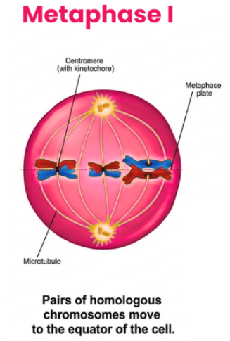
Key Features
Homologous pairs line up together
Each homologue is attached to spindle fibers from opposite poles
Orientation of each pair is random
This random arrangement is called independent assortment. It is another major source of genetic variation because maternal and paternal homologues can move into either daughter cell in many possible combinations.
Anaphase I
In Anaphase I, homologous chromosomes separate and move toward opposite poles.
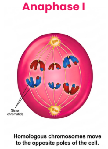
Important Point
Homologues separate
Sister chromatids do not separate yet
This is the reduction step of meiosis. Because homologous chromosomes are separated, each pole receives only one chromosome from each homologous pair. As a result, chromosome number becomes haploid.
Telophase I
In Telophase I, chromosomes reach opposite poles.
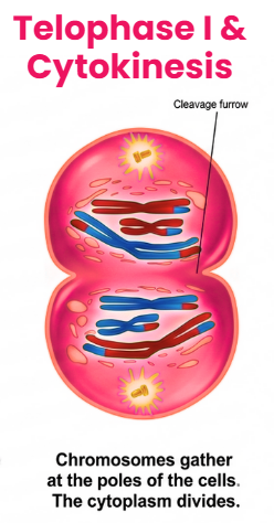
Depending on the organism:
nuclear membrane may reform
chromosomes may partly decondense
cytokinesis usually occurs
This produces two haploid daughter cells. However, each chromosome still consists of two sister chromatids, so DNA is still in duplicated form. Also, due to crossing over, sister chromatids are no longer genetically identical.
Meiosis II
Meiosis II is the second meiotic division. It is often described as similar to mitosis, but it occurs in haploid cells.
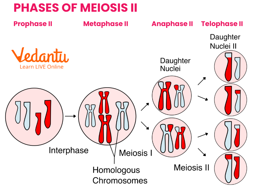
The purpose of meiosis II is to separate the sister chromatids of each chromosome. Cells do not replicate DNA before entering meiosis II.
The stages of meiosis II are:
Prophase II
Metaphase II
Anaphase II
Telophase II
Prophase II
In Prophase II:
chromosomes condense again
nuclear membrane breaks down if it had reformed
spindle fibers form
centrosomes move apart
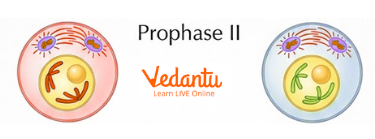
Since the cells entering meiosis II are already haploid, there is no homologous pairing here. Each chromosome is still made of two sister chromatids.
Metaphase II
In Metaphase II, chromosomes line up individually at the metaphase plate.
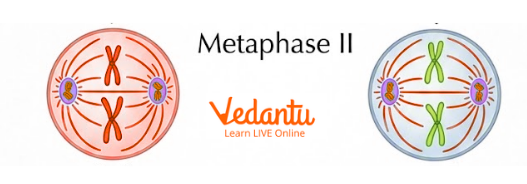
Important Feature
Unlike metaphase I, where homologous pairs lined up together, here each chromosome aligns singly. Spindle fibers attach to sister chromatids from opposite poles.
Anaphase II
In Anaphase II, the centromeres split and the sister chromatids separate. Each chromatid now becomes an independent chromosome and moves to opposite poles of the cell.
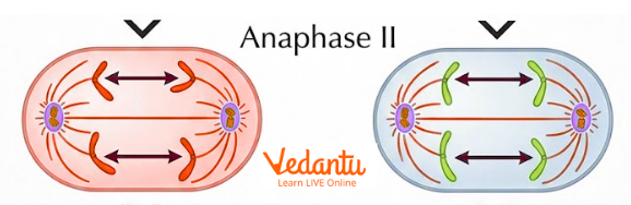
This stage resembles anaphase of mitosis, but remember that the cell is already haploid.
Telophase II
In Telophase II:
chromosomes reach the poles
nuclear membranes reform
chromosomes decondense
cytokinesis occurs
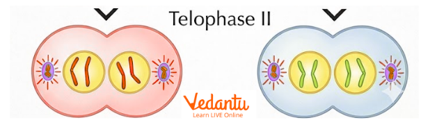
At the end of telophase II, the result is four haploid cells, each containing chromosomes with only one chromatid. In humans, these are the final gametes formed by meiosis.
Stages of Meiosis Quick Guide
Phases of Meiosis: Key Differences Between Meiosis I and Meiosis II
How Meiosis Creates Genetic Variation?
One of the most important features of meiosis is that it generates genetic diversity. The four gametes formed at the end of meiosis are not genetically identical.
The two main reasons are:
1. Crossing over
During prophase I, homologous chromosomes exchange DNA segments. Since the crossover points are more or less random, new combinations of alleles are formed.
2. Random orientation of homologous pairs
During metaphase I, homologous pairs align randomly at the equator. This causes different combinations of maternal and paternal chromosomes to enter daughter cells.
In humans, random orientation alone can produce more than 8 million different gamete combinations, and when crossing over is added, the number becomes effectively enormous.
Significance of Meiosis
1. Maintains chromosome number
Meiosis reduces chromosome number to haploid, so fertilization can restore the diploid number without doubling chromosomes in every generation.
2. Produces gametes
It forms sperm and eggs, which are necessary for sexual reproduction.
3. Creates genetic variation
Crossing over an independent assortment makes offspring genetically different from parents and from one another.
4. Supports evolution
Genetic variation is the raw material for natural selection and evolution.
5. Ensures proper distribution of chromosomes
Meiosis ensures that each gamete receives one chromosome from each homologous pair.
Thus, the significance of meiosis goes far beyond reproduction. It is also central to heredity and biodiversity.
Difference Between Mitosis and Meiosis
Meiosis in Humans
In humans:
meiosis occurs in the reproductive organs
diploid germ cells undergo meiosis
haploid gametes are formed
Chromosome Numbers
Diploid human cells: 46 chromosomes
Haploid gametes: 23 chromosomes
After fertilization:
23 from sperm + 23 from egg = 46 chromosomes in zygote
FAQs on Meiosis: Stages, Diagram, Cell Division and Significance
1. What is meiosis?
Meiosis is a type of cell division in which one diploid cell forms four haploid daughter cells, mainly for gamete formation.
2. What are the stages of meiosis?
The stages of meiosis are prophase I, metaphase I, anaphase I, telophase I, prophase II, metaphase II, anaphase II, and telophase II.
3. What happens in meiosis I?
In meiosis I, homologous chromosomes pair up, crossing over occurs, and homologues separate into two haploid cells.
4. What happens in meiosis II?
In meiosis II, sister chromatids separate, producing four haploid cells.
5. Why is meiosis called reduction division?
It is called reduction division because chromosome number is reduced from diploid to haploid.
6. What is the significance of meiosis?
Meiosis is significant because it produces gametes, maintains chromosome number, and creates genetic variation.
7. What does a meiosis diagram show?
A meiosis diagram shows homologous pairing, crossing over, chromosome separation in meiosis I, chromatid separation in meiosis II, and formation of four haploid cells.