


What is the Human Ear? Structure, Parts and Functions Explained
The human ear is a paired sensory organ present on both sides of the head. It performs two major functions that are essential for daily life: hearing and balance. In simple words, the ear detects sound waves from the surroundings and also helps the body maintain equilibrium while standing, walking, running, or changing head position. This makes the ear one of the most important sense organs in the human body.
Structure of Human Ear
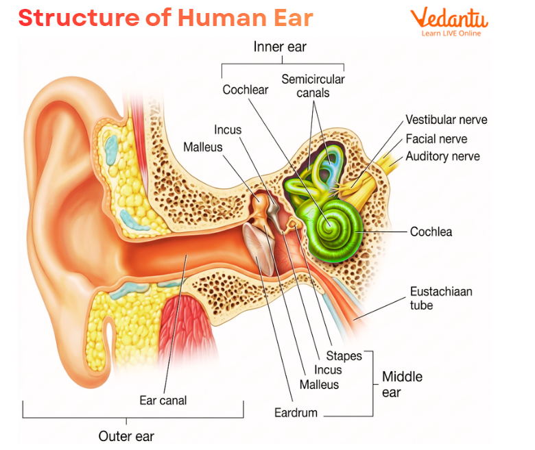
The structure of human ear is divided into three major parts:
outer ear
middle ear
inner ear
These three parts work together in a sequence. The outer ear collects sound waves. The middle ear transmits and amplifies the vibrations. The inner ear converts these vibrations into nerve impulses and also contains the organs for balance. The eardrum or tympanic membrane separates the outer ear from the middle ear.
Parts of Human Ear
1. Outer Ear
2. Middle Ear
3. Inner Ear
Each part has distinct structures and functions, but all are connected functionally to help in hearing and equilibrium.
Outer Ear
The outer ear is the visible part of the ear. It is also called the external ear, auricle, or pinna. It is made of cartilage covered by skin and has ridges and folds that help collect sound waves from the environment. The outer ear also contains glands that produce earwax.
Main Structures of the Outer Ear
auricle or pinna
external auditory canal or ear canal
earwax-secreting glands
Functions of the Outer Ear
The outer ear mainly performs the following functions:
collects sound waves from the surroundings
directs sound into the ear canal
protects the deeper ear structures
helps trap dust and foreign particles with the help of earwax
The funnel-shaped ear canal carries sound waves inward toward the tympanic membrane or eardrum. This is why the outer ear is often described as the sound-collecting part of the auditory system.
Middle Ear
The middle ear begins just beyond the eardrum. It is an air-filled chamber that contains three very small bones called the ossicles. These are:
malleus
incus
stapes
These bones are the smallest bones in the human body and are extremely important for hearing because they transmit and amplify sound vibrations from the eardrum to the inner ear.
Ossicles of the Middle Ear
1. Malleus
The malleus is attached to the tympanic membrane and receives vibrations from it.
2. Incus
The incus lies between the malleus and stapes and helps pass vibrations forward.
3. Stapes
The stapes is the last ossicle and transmits vibrations to the inner ear.
4. Eustachian Tube
The middle ear also contains the eustachian tube, which connects the middle ear to the throat. Its main job is to equalise the air pressure on both sides of the eardrum. When a person yawns, sneezes, or swallows, the eustachian tube opens and helps balance ear pressure.
Functions of the Middle Ear
receives vibrations from the eardrum
amplifies sound with the help of ossicles
transmits sound to the inner ear
maintains pressure balance through the eustachian tube
The middle ear is therefore the sound-conducting and sound-amplifying region of the auditory system.
Inner Ear
The inner ear is the most specialised part of the ear. It contains structures for both hearing and balance. The two main components of the inner ear are:
cochlea
semicircular canals
These structures are filled with fluid and contain delicate sensory receptors.
Cochlea
The cochlea is the hearing organ. It is a spiral or snail-shaped structure with fluid-filled chambers lined by tiny hair-like sensory cells. When sound vibrations reach the cochlea, the fluid inside it moves, and this movement bends the hair cells. These hair cells convert mechanical vibrations into electrical nerve impulses, which are then sent to the brain.
So, the cochlea is the place where sound vibrations are finally converted into neural signals.
Semicircular Canals
The semicircular canals are responsible for balance. These loop-shaped canals are filled with fluid and contain sensory receptors. When the head moves, the fluid inside also moves. This stimulates the sensory hairs, which send information to the brain through the vestibular nerve. The brain then coordinates with muscles to maintain posture and equilibrium.
Functions of the Inner Ear
converts sound vibrations into electrical signals
sends auditory information to the brain
detects head movement
helps maintain balance and body orientation
This is why damage to the inner ear may affect both hearing and balance. Always remember the sound pathway:
outer ear → ear canal → eardrum → ossicles → inner ear → cochlea → nerve impulses → brain
At the same time, the semicircular canals are shown as the balance-related structures of the inner ear.
Function of the Human Ear
The human ear has two major functions:
1. Hearing
2. Balance
These two roles are closely linked to the structure of the ear.
Hearing Function
When sound waves enter the ear canal, they strike the eardrum and make it vibrate. These vibrations are carried through the ossicles and then to the cochlea. Inside the cochlea, the sensory hair cells convert these vibrations into electrical signals that are carried to the brain. The brain interprets these signals as sound.
Balance Function
The inner ear also helps maintain balance. The semicircular canals contain fluid and sensory hairs. When the head moves, the fluid shifts and stimulates these hairs. The information is sent through the vestibular nerve to the brain, which then controls muscles and posture to keep the body balanced.
So, the ear is not only an organ of hearing. It is also an organ of equilibrium.
Mechanism of Hearing
The process of hearing can be explained step by step.
Step 1: Sound Collection
The pinna collects sound waves and directs them into the ear canal.
Step 2: Eardrum Vibration
The sound waves strike the tympanic membrane, causing it to vibrate.
Step 3: Ossicle Movement
The malleus, incus, and stapes receive these vibrations and amplify them.
Step 4: Transfer to the Inner Ear
The amplified vibrations reach the inner ear.
Step 5: Cochlear Fluid Movement
Inside the cochlea, the fluid moves in response to the vibrations.
Step 6: Hair Cell Stimulation
Tiny hair cells in the cochlea bend and convert the vibrations into electrical impulses.
Step 7: Brain Interpretation
These impulses travel to the brain, where they are interpreted as sound.
Mechanism of Balance
The ear also contributes to equilibrium through the vestibular apparatus.
How Balance is Maintained?
the semicircular canals are filled with fluid
head movement shifts this fluid
hair-like sensors detect the movement
signals are sent through the vestibular nerve
the brain receives the information and adjusts muscle activity accordingly
This is how the ear helps maintain body posture and coordination.
Audible Range of Human Ear
The audible range of the human ear refers to the range of sound frequencies that a normal human ear can hear.
The normal audible range of human ear is approximately:
20 Hz to 20,000 Hz
1. Infrasonic Sound
Frequencies below 20 Hz are called infrasonic and are not heard by humans.
2. Ultrasonic Sound
Frequencies above 20,000 Hz are called ultrasonic and are also beyond the hearing range of humans.
Common Ear Problems
Many diseases and disorders can affect the ears. Some major ear conditions include:
ear infection or otitis media
eustachian tube dysfunction
swimmer’s ear or otitis externa
ruptured eardrum
otosclerosis
perichondritis
vestibular neuritis
Meniere’s disease
ear injuries
ear tumours
Ear Infection
Often occurs in the middle ear when bacteria or viruses get trapped.
Eustachian Tube Dysfunction
Occurs when the tube is blocked, causing fullness, tinnitus, or muffled hearing.
Swimmer’s Ear
An infection of the ear canal, often caused by bacteria or fungi after water exposure.
Ruptured Eardrum
A tear or hole in the tympanic membrane due to infection, trauma, loud noise, or foreign object.
Otosclerosis
Abnormal bone remodelling in the middle ear that hardens ossicles and affects hearing.
Vestibular Neuritis
Inflammation of the vestibular nerve causing vertigo, nausea, and balance issues.
Meniere’s Disease
A chronic inner ear disorder causing dizziness, vertigo, and a feeling of fullness in the ear.
Symptoms of Ear Disorders
Common warning signs of ear problems include:
ear pain
infection
clogged ears
muffled hearing
itchy ears
nausea and vomiting
feeling of fullness in the ears
ear drainage
These symptoms should not be ignored, especially if hearing loss or dizziness is present.
Tests Used to Check Ear Function
Doctors use several tests to examine the ears and assess hearing function.
Pure-Tone Testing
Measures the quietest sounds a person can hear at different frequencies.
Middle Ear Tests
Check the function of the eardrum and middle ear.
Speech Testing
Assesses how well a person hears and repeats spoken words.
Auditory Brainstem Response (ABR)
Measures how the brain responds to sound.
Otoacoustic Emissions (OAEs)
Checks how well the cochlea is functioning by measuring sound responses from the inner ear.
Ear Care and Prevention
Good ear care helps preserve both hearing and balance. Important ear-care habits include:
keep ears dry while swimming
avoid inserting cotton swabs into the ear canal
use protective gear during contact sports
keep headphone volume low
use earplugs around loud sounds
go for routine ear examinations
These simple habits can reduce the risk of infection, injury, and hearing damage.
FAQs on Human Ear: Structure, Parts, Diagram, Functions, Ear Problems and Audible Range
1. What are the 3 main parts of the ear?
The 3 main parts of the ear are:
Outer ear
Middle ear
Inner ear
The outer ear collects sound, the middle ear passes and amplifies vibrations, and the inner ear helps with hearing and balance.
2. What is the most painful ear infection?
Otitis externa, also called swimmer’s ear, is often one of the most painful ear infections. It can cause severe ear pain, especially when the ear is touched or pulled.
3. What are the first signs of inner ear issues?
The first signs of inner ear problems often include:
Dizziness
Vertigo or spinning sensation
Loss of balance
Nausea or vomiting
Hearing loss
Tinnitus or ringing in the ear
4. What is the location of the ear?
The ear is located on both sides of the head. It lies over the temporal region and is placed slightly behind the jaw area.
5. Which part of the ear is most important?
The inner ear is often considered the most important part because it contains the cochlea for hearing and the semicircular canals for balance.
6. What causes ear pain?
Common causes of ear pain include:
Ear infection
Wax buildup
Eustachian tube blockage
Pressure changes
Injury
Jaw or throat problems
7. What is the best treatment for ear infection?
The best treatment depends on the cause. Common treatment includes:
Pain relief medicines
Warm compress
Antibiotics if the infection is bacterial
Medical check-up if symptoms are severe or do not improve
8. What is stage 3 ear infection?
Stage 3 ear infection usually means chronic otitis media, where fluid stays in the middle ear for a long time and may lead to hearing problems if not treated.
9. What are the five common symptoms of ear disease?
Five common symptoms of ear disease are:
Ear pain
Hearing loss
Fluid coming from the ear
Swelling around the ear
Feeling unwell or fever
10. Which organ is connected to the ear?
The ear is connected to the throat by the Eustachian tube and to the brain by the auditory and vestibular nerves.
11. What is the three finger test for ear?
The three finger test for ear is a physical check used to look for mastoid tenderness or infection behind the ear. Pressure is applied to specific points around the ear to see if pain is present.