

How Does the Human Ear Work? Step-by-Step Explanation
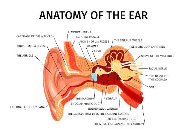
The human ear is a remarkable organ responsible for detecting, transmitting, and transducing sound. In addition to hearing, it plays a vital role in maintaining balance. This guide will help you understand the complete structure of ear anatomy and the function of the ear in an easy-to-understand manner. We have included an interactive ear structure diagram and a structure of ear labelled illustration to aid visual learning.
The human ear is a sensitive and sophisticated organ. Its primary responsibilities include receiving sound waves, converting them into neural signals, and maintaining balance. Understanding the structure of ear anatomy is crucial for appreciating how the ear works and how its internal structure of the ear supports both auditory and balance functions.
Also Check: Sense Organs
Detailed Structure of Ear Anatomy
The human ear is divided into three main sections, each with a specific role. Below is a detailed breakdown of the structure of ear anatomy along with a description that will help you visualise an accurate ear structure diagram.
External Ear
The external ear consists of:
Auricle (Pinna): A thin, curved plate of elastic cartilage covered by skin. Its funnel-like curves collect sound waves and direct them towards the ear canal. This part is visible in the structure of ear-labelled diagrams.
External Auditory Meatus: A slightly curved canal that is part bony and part cartilaginous. It is lined with stratified epithelium and contains wax glands which help in trapping debris.
Tympanic Membrane: Commonly known as the eardrum, it vibrates when sound waves hit it, passing the vibrations onto the middle ear.
Middle Ear
The middle ear acts as a bridge between the external and internal ear:
Tympanic Cavity: An air-filled space separated by the tympanic membrane from the external ear and by a bony wall from the internal ear.
Eustachian Tube: A 4cm long tube that connects the tympanic cavity to the nasopharynx. Its role is to equalise air pressure on both sides of the tympanic membrane.
Ear Ossicles: Three small bones—malleus (hammer), incus (anvil), and stapes (stirrup)—transmit sound vibrations from the tympanic membrane to the inner ear. These tiny bones are a crucial part of the structure of ear anatomy and are often highlighted in detailed ear structure diagram illustrations.
Internal Ear
The internal ear is where sound is converted into electrical signals and where balance is regulated:
Bony Labyrinth: Contains the vestibule, three semi-circular canals, and the coiled cochlea. This structure is filled with perilymph fluid.
Membranous Labyrinth: Residing within the bony labyrinth, it is filled with endolymph and includes the semi-circular ducts, cochlear duct, saccule, and utricle. Sensory receptors, such as those in the organ of Corti, are located here and play a major role in hearing.
Function of the Ear
The function of the ear involves two key processes: hearing and balance.
Hearing
Sound Collection: Sound waves are gathered by the auricle and travel through the external auditory meatus.
Vibration Transmission: The tympanic membrane vibrates when sound waves hit it, and these vibrations are transferred through the ear ossicles.
Signal Conversion: The stapes transmit the vibrations to the inner ear’s cochlea, where the vibrations are converted into electrical signals by the hair cells in the organ of Corti.
Signal Processing: These electrical signals travel via the auditory nerve to the brain, where they are interpreted as sound.
Balance
Equalisation of Pressure: The Eustachian tube plays a crucial role by equalising air pressure in the middle ear.
Balance Maintenance:The vestibular system in the internal ear, which includes the semi-circular canals and the otolithic organs (saccule and utricle), sends signals to the brain to help maintain balance and spatial orientation.
Glossary
Auricle (Pinna): The outer part of the ear that collects sound.
Tympanic Membrane: The eardrum that vibrates to transmit sound.
Ossicles: The three small bones (malleus, incus, stapes) that convey sound vibrations.
Cochlea: A spiral-shaped organ responsible for converting sound waves into nerve impulses.
Vestibular System: The components within the inner ear that help regulate balance.
Related Links:


FAQs on Human Ear: Structure, Function, and Detailed Anatomy Guide
1. What are the two primary functions of the human ear?
The human ear is a complex organ with two main jobs. Its primary function is hearing, which involves detecting sound waves, amplifying them, and converting them into electrical signals for the brain to interpret. Its second crucial function is maintaining balance or equilibrium, managed by the vestibular system located in the inner ear.
2. Can you explain the main sections of the human ear?
The human ear is divided into three main sections, each with a specific role:
- The Outer Ear: This includes the visible part called the pinna and the ear canal. Its job is to collect sound waves and channel them towards the eardrum.
- The Middle Ear: This is an air-filled cavity that contains three tiny bones called ossicles (malleus, incus, and stapes). It transmits and amplifies the sound vibrations from the eardrum to the inner ear.
- The Inner Ear: This fluid-filled section contains the cochlea for hearing and the vestibular system for balance. It converts mechanical vibrations into nerve impulses.
3. What is the specific role of the ear ossicles in the process of hearing?
The ear ossicles—the malleus (hammer), incus (anvil), and stapes (stirrup)—form a tiny bridge connecting the eardrum to the inner ear. Their main role is to act as a lever system that amplifies the sound vibrations. This amplification is necessary to transfer the energy from the air-filled middle ear to the fluid-filled inner ear efficiently, ensuring the sound signal is strong enough to be processed.
4. How does the cochlea in the inner ear help us hear different sounds?
The cochlea is a spiral-shaped, fluid-filled organ lined with thousands of tiny hair cells. When vibrations from the middle ear reach the cochlea, they create waves in the fluid. Different frequencies (or pitches) of sound cause different parts of the cochlea's membrane to vibrate. High-pitched sounds are detected at the base, while low-pitched sounds are detected at the apex. These vibrations stimulate the hair cells, which then generate electrical signals sent to the brain.
5. How is our sense of balance connected to the structure of the inner ear?
The sense of balance is managed by the vestibular system in the inner ear, which is separate from the cochlea. It consists of the semicircular canals and the otolith organs. The semicircular canals detect rotational movements of the head (like nodding or shaking), while the otoliths detect linear acceleration and gravity (like when you're in a moving car). Information from this system is sent to the brain, helping it understand the body's position and maintain stability.
6. Why is the inner ear filled with fluid instead of air like the middle ear?
The fluid in the inner ear, known as perilymph and endolymph, is essential for converting sound vibrations into a format the nervous system can understand. Sound travels differently through fluid than through air. The fluid medium allows the creation of precise pressure waves within the cochlea that stimulate specific hair cells according to the sound's pitch. This fine-tuned mechanism would not be possible with air, which is less effective at transmitting these detailed vibrations to the sensory cells.
7. What is the importance of the Eustachian tube for ear function?
The Eustachian tube is a small passageway that connects the middle ear to the back of the throat. Its main function is to equalise the air pressure on both sides of the eardrum. This is why your ears might 'pop' during a flight or when changing altitude. By ensuring the pressure is balanced, it allows the eardrum to vibrate freely and correctly, which is vital for clear hearing.
8. What happens after the ear converts sound into electrical signals?
Once the sensory hair cells in the cochlea convert sound vibrations into electrical signals, these signals travel along the auditory nerve to the brain. They reach the auditory cortex in the brain's temporal lobe, which is responsible for processing them. The brain then interprets these signals, allowing us to perceive them as distinct sounds, words, or music and to identify their direction, volume, and pitch.


