

What Is the Structure and Function of the Human Heart
The heart is a muscular organ. This organ circulates blood through the circulatory system's blood vessels. Pumped blood transports oxygen and nutrients to the body while transporting metabolic waste like carbon dioxide to the lungs. In humans, the heart is about the size of a closed fist and is located in the middle compartment of the chest, between the lungs.
Every day, our heart does amazing things. Every cell in the human body, except the cornea, receives blood from the heart. In the United States, heart disease is the leading cause of death. That is why it is critical to take care of the heart by living a heart-healthy lifestyle.
The Human Heart
The heart is a small organ that circulates blood throughout our body. It is our circulatory system's primary organ. The heart functions as two pumps, one on each side, which work in tandem. Blood flows from the right atrium to the right ventricle, then to the lungs to be oxygenated. Blood flows from the lungs to the left atrium, then to the left ventricle. Our heart's primary function is to keep oxygenated blood circulating throughout our body.
Structure of the Heart
As it can be seen in the structure of the heart diagram, our heart is composed of four chambers: two small upper chambers called atria and two bigger bottom chambers called ventricles.
The ventricle walls are significantly thicker than those of the atria.
The interatrial septum is a thin, muscular wall that separates the right and left atria, whereas the interventricular septum is a thick-walled wall that separates the right and left ventricles.
The atrioventricular septum is a strong fibrous structure that divides the atrium and ventricle of the same side. However, each atrioventricular septum has an opening through which the two chambers on the same side are joined.
The chordae tendineae are unique fibrous cords that are linked to the flaps of the bicuspid and tricuspid valves at one end and to the ventricular wall at the other end, with specific muscles termed papillary muscles.
Three semilunar valves are present, where the pulmonary artery (which arises from the right ventricle and transports deoxygenated blood to the lungs) and aorta (which arises from the left ventricle and transports oxygenated blood to other regions of the body) leave the heart. These valves keep blood from returning to the ventricles.
Deoxygenated blood enters the right atrium via the coronary sinus and two big veins known as vena cava as shown in the image of a labelled diagram. Through two pairs of pulmonary veins, the left atrium gets oxygenated blood from the lungs.
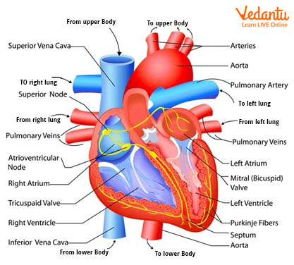
Well Labelled Diagram of The Heart
Right atrium, right ventricle, left atrium, left ventricle, tricuspid valve, mitral valve, pulmonary valve, aortic valve, superior vena cava, inferior vena cava, pulmonary trunk, right pulmonary artery, left pulmonary artery, pulmonary veins, and aorta etc. form the main part of the heart.
Layers of The Human Heart
Pericardium: Pericardium is the membrane that surrounds and protects the heart. It limits the heart to its mediastinum location while providing enough mobility for robust and fast contraction.
Fibrous Pericardium: Tough, inelastic, thick, and uneven connective tissue. It protects the heart and anchors it in the mediastinum by preventing overstretching.
Serous Pericardium: It is a thinner, more sensitive membrane that forms a second layer surrounding the heart. The fibrous pericardium is linked to the outer parietal layer. The inner visceral layer, also known as the epicardium, is one of the layers of the heart wall that adheres tightly to the surface of the heart. The pericardial fluid is found in the area between the parietal and visceral layers, and the region that contains this lubricating secretion of pericardial cells is known as the pericardial cavity.
The heart's wall is made up of three layers: the epicardium (external layer), the myocardium (middle layer), and the endocardium (inner layer).
Epicardium: It is the clear outermost layer. It is made up of mesothelium and fragile connective tissue, which gives the surface heart a smooth, slippery touch.
Myocardium: The middle layer of cardiac muscle tissue that makes up around 95 per cent of the heart. It is responsible for its pumping activity. The cardiac muscle is an involuntary muscle.
Endocardium: The deepest layer, is a thin layer of endothelium that is covered by a thin layer of connective tissue. It creates a smooth lining for the heart chambers and valves. It is continuous with the endothelial lining of big blood arteries connected to hearts and reduces surface friction as blood flows through the heart and blood vessels.
Chambers of The Heart
The heart is made up of four chambers, as seen in the labelled diagram of the heart. The atria (entrance halls or chambers) are the two superior receiving chambers, while the ventricles (little bellies) are the two lower pumping chambers. An auricle is a wrinkled, pouch-like structure on the front surface of each atrium.
1. Right Atrium:
The right atrium is located on the heart's right side and receives blood from three veins: the superior vena cava, inferior vena cava, and coronary sinus.
The posterior wall is smooth, but the anterior wall is rough due to the presence of pectinate muscles, which extend the auricle.
The interatrial septum is a thin partition between the right and left atriums.
2. Right Ventricle:
The right ventricle is roughly 4-5 mm thick on average and makes up the majority of the heart's anterior surface.
The right ventricle is divided from the left ventricle by an internal barrier known as the interventricular septum.
The pulmonary valve (pulmonary semilunar valve) directs blood from the right ventricle into the pulmonary trunk, which separates into right and left pulmonary arteries.
Arteries are usually responsible for transporting blood away from the heart.
3. Left Atrium:
The left atrium is roughly the same thickness as the right atrium and makes up the majority of the heart's base.
It gets blood from the lungs via four pulmonary veins.
The interior of the left atrium, like the right atrium, has a smooth posterior wall.
Since pectinate muscles are restricted to the left atrial auricle, the anterior wall of the left atrium is likewise smooth.
The bicuspid valve, which has two cusps, allows blood to flow from the left atrium into the left ventricle.
4. Left Ventricle:
The left ventricle is the thickest chamber of the heart, usually 10-15 mm in thickness, and serves as the heart's apex.
The left ventricle, like the right, features trabeculae carneae and chordae tendineae that connect the bicuspid valve cusps to papillary muscles.
Blood flows from the left ventricle into the ascending aorta via the aortic valve.
Functions of The Heart
The cardiac cycle, which is the heart's blood-pumping cycle, ensures that blood is dispersed throughout the body.
The oxygen distribution process begins when oxygen-free blood enters the heart through the right atrium, travels to the right ventricle, enters the lungs for oxygen replenishment and carbon dioxide release, and then returns to the left chambers for redistribution.
When a cardiovascular condition is detected, the heart's function can be checked. A heart-related ailment, for example, is characterised by a consistently irregular heartbeat or beats per minute. This is because a heartbeat is a representation of the heart's two-phase oxygen-reloading mechanism.
Conclusion
Every minute of a 24-hour day, our heart beats anywhere from 60 to 100 times. It beats roughly 100,000 times per day. But, unlike the other muscles in our bodies, our heart almost never gets tired until it stops completely. Our heart does amazing things, and we have studied the structure of the heart diagram with parts and different muscles which form the heart. This article provides important information about its functioning. Our heart is helping us stay healthy, we must take care of it. From an exam point of view, one must practise heart structure with labelling. Human heart diagram and functions are frequently asked in the examination.


FAQs on Heart Anatomy and Blood Circulation in Humans
1. What is the heart and what does it do?
The heart is a muscular organ that pumps blood throughout the body to supply oxygen and nutrients and remove wastes. It works as the central organ of the circulatory system.
- Pumps oxygenated blood to body tissues
- Receives deoxygenated blood from the body
- Maintains blood pressure and circulation
2. Where is the heart located in the human body?
The heart is located in the chest cavity between the lungs, slightly to the left of the midline. It lies in a space called the mediastinum.
- Positioned behind the sternum (breastbone)
- Above the diaphragm
- Protected by the rib cage
3. What are the main parts of the heart?
The human heart has four chambers and associated valves and vessels. The main parts include:
- Right atrium – receives deoxygenated blood
- Right ventricle – pumps blood to the lungs
- Left atrium – receives oxygenated blood
- Left ventricle – pumps blood to the body
4. How does the heart pump blood step by step?
The heart pumps blood through a coordinated cycle called the cardiac cycle. The steps are:
- Deoxygenated blood enters the right atrium
- Blood moves to the right ventricle and is pumped to the lungs
- Oxygenated blood returns to the left atrium
- Blood flows into the left ventricle and is pumped to the body
5. What is the function of heart valves?
The heart valves ensure one-way blood flow through the heart by preventing backflow. The four main valves are:
- Tricuspid valve
- Pulmonary valve
- Mitral (bicuspid) valve
- Aortic valve
6. What is the difference between arteries and veins in heart circulation?
The main difference is that arteries carry blood away from the heart, while veins carry blood toward the heart. In heart circulation:
- Arteries usually carry oxygenated blood (except the pulmonary artery)
- Veins usually carry deoxygenated blood (except the pulmonary veins)
- Arteries have thicker walls than veins
7. What controls the heartbeat?
The heartbeat is controlled by a natural pacemaker called the sinoatrial (SA) node. It generates electrical impulses that regulate heart rhythm.
- SA node initiates the electrical signal
- Signal spreads to the atrioventricular (AV) node
- Impulses travel through specialized fibers to ventricles
8. What is the cardiac cycle?
The cardiac cycle is the sequence of events that occurs during one complete heartbeat. It consists of:
- Systole – contraction phase when blood is pumped out
- Diastole – relaxation phase when chambers fill with blood
9. Why is the left ventricle thicker than the right ventricle?
The left ventricle has a thicker muscular wall because it pumps blood to the entire body under high pressure. In contrast:
- The right ventricle pumps blood only to the lungs
- Systemic circulation requires more force than pulmonary circulation
10. What is coronary circulation?
The coronary circulation is the blood supply that delivers oxygen and nutrients directly to the heart muscle itself. It involves:
- Coronary arteries branching from the aorta
- Capillary networks within the heart muscle
- Cardiac veins that return blood to the right atrium


