

Labeled diagram of human eye with parts and functions
The Human Eye is the main visual organ of our human body. It is just like a camera having a lens in it. There are different colors of human eyes. Have you noticed it? This is specially due to the color of eyes. There are many things that are in the eye. Even the formation of an image includes a number of processes. All this information you are going to receive in this article in brief. Human Eye due to not being taken care of properly results in getting defected and you will know about such defects here also. Firstly let's have a look at the human eye.
The human eye is one of the most important organs of the human body which when interacted with light gives us the sense of sight or vision. There are two kinds of cells in the eye namely rods and cones. The basic functions of Rods and Cones are conscious light perception, color differentiation and depth perception. The human eye is capable of distinguishing between about 10 million colors, and it can also detect a single photo. The human eye is a part of the sensory nervous system.
Labeled Diagram of Human Eye
The eyes of all mammals consist of a non-image-forming photosensitive ganglion within the retina which receives light, adjusts the dimensions of the pupil, regulates the availability of melatonin hormones, and also entertains the body clock.
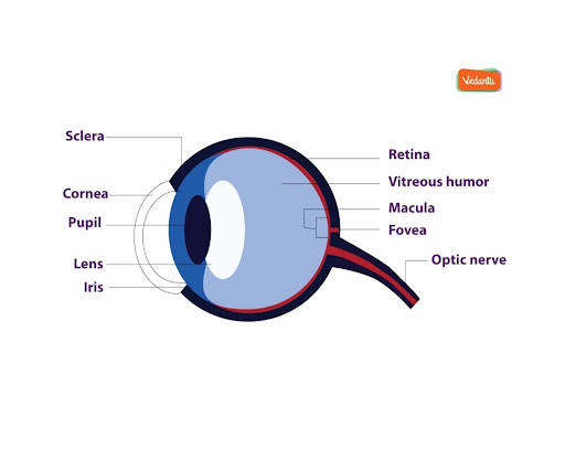
The anterior chamber of the eyes is the space between the cornea and therefore the iris and is crammed with a lubricating fluid, aqueous humor.
The vascular layer of the eye , referred to as the choroid, contains the animal tissue . The vascular layer consists of blood vessels. This layer is filled with blood, that is this layer has a good supply of blood.
The iris and therefore the choroid are connected by the membrane . The iris is a colored part of the eye that provides color to your eyes. The color of the eye of any person you witness is due to the iris part of their eye.
Fovea is a minute pit placed in the macula of the retina that helps in forming a clear vision. Fovea is a pit structure that is present in the layer macula lutea. This pit is situated in the posterior part of the eyeball.
Cornea is a dome-shaped tissue covering the front of the eye. This dome-shaped cornea is a transparent dome that will help the light to pass through them up to the lens,resulting in the formation of the image at the back of the eye,that is the retina.
Iris is that it coloured a part of the eyes and controls the quantity of light entering the eyes by regulating the dimensions of the pupil.
The lens is located just behind the iris. Its function is to focus the light on the retina. Lens presence also divides the front part of the eye into anterior and posterior chamber. This focuses the light rays falling onto it on the retina causing the formation of the image.
The optic nerve helps in transmitting the electrical signals from the retina to the brain. The optic nerve arises from the optic disc and it takes the information of any object or image that you are seeing up to the brain.
Pupil is the opening at the center of the iris. Its size changes with the amount of light. Pupil dilates. I'm the dim light and it constricts in the bright light causing the right amount of light to enter the eye, thus forming an image on the retina.
The retina lines contain several photoreceptors. These photoreceptors are the ones that can absorb light and prevent the scattering of the light.
Vitreous humor is the fluid present within the center of the attention and provides form and shape to the eyes. This is the posterior chamber of the eye. The anterior chamber of the eye is filled with aqueous humor that helps to provide shape to the anterior portion of the eye.
Operation of an Human Eye
The operation of a human eye can be compared to that of a digital camera because of the following reasons -
Light focuses mainly on the cornea, which acts sort of like an optical lens .
The iris controls the light that reaches by adjusting the dimensions of the pupil, and thus it functions just like the diaphragm of a camera.
The lens of attention is found behind the pupil, and it focuses light. This lens helps the attention to automatically specialize in near and distant objects, and also the approaching objects, like an autofocus optical lens .
The cornea and lens focus light to succeed in the retina, which may be a light-sensitive zone present on the inner lining of the rear of the eye .
The retina converts optical phenomenon images into electronic signals, and thus it acts as a bitmap sensor of a camera . These electric signals are then transmitted by the nervous opticus to the visual area , which is liable for the sense of sight.
Function of the Human Eye
Human eyes are the organs that will help you to see. This provides you with a sense of vision. Our eyes contain visual receptors. Anything that you will see is just because of the presence of these visual receptors. Any object you see , the light rays first come through the cornea. The cornea is a transparent layer that will help you to pass the light upto the lens. There is iris present in the eyes that will provide you with the color. The light rays that enter up to the lens are focused back on the retina, where the inverted image is formed. This inverted image is then formed directly in our brain. Whichever image we see, the direct image is formed in the mind before the inverted image is formed on the retina.Human eyes are a specialized sense organ capable of receiving visual images and producing the sense of sight in us. The eye receives direct oxygen through the aqueous humor. The aqueous humor nourishes the cornea, lens, and therefore the iris, by carrying nutrients, removing waste materials excreted by the lens, and maintaining the form and structure of the eyes. The aqueous humor is liable for providing shape to the eyes. It must be clear to function properly.
Function of Lens in the Human Eye
The main function of this lens is to focus the light rays that come into our eyes.The lens may be a transparent flexible tissue located directly behind the iris and therefore the pupil. To focus light and images on the retina becomes the basic function of the lens. The cornea and the lens are responsible for focusing the image in the retina.
Due to the elasticity & flexibility the lens has, it can change its curved shape to focus on nearby or distant objects as per the requirements. The lens provides around 25-35 you look after the entire focusing power of the eyes.
The lens is attached to the ciliary muscles, which contracts and releases in order to change the shape of the lens and also its curvature.
Due to the defect in the lens certain conditions may arise; they are termed myopia, hypermetropia, cataract and so on. Hypermetropia and myopia both can be cured by the particular kind of lens that you will use to clear the defect and there are conditions where due to old age the visual power of the lens constantly decreases.


FAQs on Human Eye Diagram and Detailed Explanation
1. What is a diagram of the eye?
A diagram of the eye is a labeled illustration that shows the structure and parts of the human eye and how they are arranged. It helps learners understand the anatomy and function of different components involved in vision.
- Shows external and internal structures
- Labels parts such as cornea, retina, and optic nerve
- Explains how light travels through the eye
- Used in Biology exams and practical learning
2. What are the main parts labeled in a diagram of the human eye?
The main parts labeled in a human eye diagram include structures that help in vision and image formation. These parts work together to detect and process light.
- Cornea – transparent front layer
- Iris – colored part controlling pupil size
- Pupil – opening that allows light entry
- Lens – focuses light on the retina
- Retina – light-sensitive inner layer
- Optic nerve – carries signals to the brain
- Sclera – white protective outer layer
3. What is the function of the retina in the eye diagram?
The retina is the light-sensitive layer of the eye that converts light into nerve impulses. It contains specialized photoreceptor cells that detect light and color.
- Rods – detect dim light and black-and-white vision
- Cones – detect color and bright light
- Transmits signals through the optic nerve to the brain
4. How does light travel through the eye according to the diagram?
Light travels through specific structures of the eye in a fixed sequence to form an image on the retina. This pathway ensures proper focusing and image formation.
- Light enters through the cornea
- Passes through the pupil
- Gets focused by the lens
- Forms an image on the retina
- Signals travel via the optic nerve to the brain
5. What is the function of the lens in the eye?
The lens focuses light rays onto the retina to form a clear image. It changes its shape to focus on objects at different distances, a process called accommodation.
- Becomes thicker to see nearby objects
- Becomes thinner to see distant objects
- Works with ciliary muscles to adjust focus
6. What is the difference between rods and cones in the retina?
The main difference between rods and cones is that rods detect dim light while cones detect color and bright light. Both are photoreceptors located in the retina.
- Rods: Night vision, sensitive to low light, no color detection
- Cones: Daylight vision, detect red, green, and blue colors
- Cones are concentrated in the fovea
7. What is the role of the optic nerve in the eye diagram?
The optic nerve carries visual nerve impulses from the retina to the brain. It connects the eye to the visual centers in the cerebrum.
- Transmits electrical signals generated by photoreceptors
- Enters the brain at the optic chiasma
- Essential for interpretation of images
8. What is the function of the iris and pupil?
The iris controls the size of the pupil, and the pupil regulates the amount of light entering the eye. Together, they adjust to different light conditions.
- In bright light: pupil constricts
- In dim light: pupil dilates
- Helps protect the retina from excessive light
9. Why is the cornea important in the structure of the eye?
The cornea is important because it is the first surface that refracts (bends) light entering the eye. It plays a major role in focusing vision.
- Transparent and dome-shaped
- Provides most of the eye’s refractive power
- Protects internal eye structures
10. How do you draw and label a diagram of the human eye?
To draw and label a diagram of the human eye, sketch the basic outline and clearly mark the internal structures in correct positions. A neat and proportionate diagram is important for exams.
- Draw a circular outline for the eyeball
- Add the cornea at the front
- Draw the iris and pupil
- Sketch the lens behind the pupil
- Mark the retina at the inner back layer
- Label the optic nerve extending from the rear


