

What are the phases of the cardiac cycle with diagram and explanation
The human heart is a muscular organ, roughly the size of a fist, responsible for pumping blood through the body. It plays a vital role in delivering oxygen and nutrients, while also removing waste products from tissues. The movement of blood within the cardiovascular system is made possible by the intricate processes of the cardiac cycle.
Also Read: Structure and Function of the Human Heart
What is the Cardiac Cycle?
The cardiac cycle refers to the complete sequence of events that occur during one heartbeat, including both the contraction and relaxation phases of the heart chambers. The cycle ensures that blood is pumped effectively to the lungs and the rest of the body. A typical cardiac cycle is divided into several phases, where the heart muscles contract and relax in a coordinated way, allowing blood to flow through the body efficiently.
Read More: Blood Cells
Cardiac Cycle in Numbers: A healthy heart beats around 72 times per minute, meaning that one full cardiac cycle occurs approximately 72 times in a minute. This is equivalent to around 0.8 seconds per cycle. The cycle consists of both systolic (contraction) and diastolic (relaxation) phases.
Step-by-Step Cardiac Cycle Flow Chart
A step-by-step cardiac cycle flow chart outlines each phase in sequence, making it easier for students to understand how the heart operates. Here's a simplified flow:
Atrial Diastole: Relaxation of atria; blood flows into the ventricles.
Atrial Systole: Atria contract, filling ventricles.
Isovolumic Contraction: Ventricles contract, valves close.
Ventricular Ejection: Blood is pumped from the ventricles to the lungs and body.
Isovolumic Relaxation: Ventricles relax, valves close to prevent backflow.
Ventricular Filling: Blood fills the ventricles from the atria.
Atrial Diastole: Rest phase before the cycle repeats.
Cardiac Cycle Diagram
A cardiac cycle diagram visually demonstrates the relationship between the phases, illustrating how blood moves through the heart’s chambers and valves. The diagram can also help show how the heart responds to electrical signals that initiate contraction and relaxation.
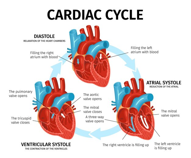
Phases of a Cardiac Cycle
There are 7 Phases of a Cardiac Cycle. Understanding these phases is essential for comprehending how the heart functions and maintains circulation throughout the body.
Atrial Diastole: The heart chambers are in a relaxed state. During this phase, the atrioventricular valves (AV valves) are open, allowing blood to flow from the atria to the ventricles. Both the aortic and pulmonary valves are closed during this time.
Atrial Systole: In this phase, the atria contract, pushing the remaining blood into the ventricles. This phase helps fill the ventricles to their maximum capacity.
Isovolumic Contraction: The ventricles start contracting, but there is no change in the volume of blood inside them yet. The AV valves close, and the aortic and pulmonary valves remain shut.
Ventricular Ejection: During this phase, the ventricles contract forcefully, and blood is pumped into the pulmonary trunk and aorta. The aortic and pulmonary valves open to allow the ejection of blood.
Isovolumic Relaxation: After the blood is ejected, the ventricles begin to relax, but no blood flows into them yet. The aortic and pulmonary valves close, preventing backflow of blood.
Ventricular Filling: Blood flows from the atria into the ventricles as the AV valves open. This is the phase where the ventricles refill with blood, ready to begin the cycle again.
Atrial Diastole: The heart enters a brief period of relaxation before the next cycle begins.
Also Read: Cardiac Output
The Function of the Cardiac Cycle
The cardiac cycle is essential for maintaining an efficient and continuous flow of blood throughout the body. It ensures that oxygen-rich blood is delivered to the organs and tissues while removing carbon dioxide and waste products. Through the rhythmic contraction and relaxation of the heart's chambers, the body maintains proper circulation, thus enabling normal bodily function.
The function of the cardiac cycle can be understood through these key tasks:
Oxygen delivery: The left ventricle pumps oxygen-rich blood through the aorta to the body.
Waste removal: The right ventricle pumps deoxygenated blood to the lungs for gas exchange (removal of carbon dioxide and replenishment of oxygen).
Blood pressure regulation: Through the heart’s pumping action, blood pressure is maintained, ensuring the proper flow to all body parts.
Duration of the Cardiac Cycle
In a healthy adult, the cardiac cycle duration is about 0.8 seconds, with the heart beating at 72 beats per minute. The exact duration of each phase varies:
Atrial Systole: ~0.1 seconds
Ventricular Systole: ~0.3 seconds
Atrial Diastole: ~0.7 seconds
Ventricular Diastole: ~0.5 seconds
The overall cycle duration remains approximately constant but can adjust depending on factors like exercise or relaxation.
Read More: Regulation of Cardiac Activity
Key Facts About the Cardiac Cycle
The cardiac cycle involves two main phases: systole (contraction) and diastole (relaxation).
The left ventricle pumps oxygen-rich blood to the body, while the right ventricle pumps deoxygenated blood to the lungs.
Electrical impulses from the sinoatrial (SA) node initiate the cycle, helping coordinate the heart's rhythm.
The cardiac cycle duration is essential in determining heart rate and overall cardiovascular health.


FAQs on Cardiac Cycle in Human Heart
1. What is the cardiac cycle?
The cardiac cycle is the sequence of events that occurs in the heart during one complete heartbeat, including contraction and relaxation of the chambers.
It consists of:
- Atrial systole – contraction of the atria
- Ventricular systole – contraction of the ventricles
- Complete cardiac diastole – relaxation of all chambers
2. What are the phases of the cardiac cycle?
The phases of the cardiac cycle are atrial systole, ventricular systole, and complete cardiac diastole.
They occur in this order:
- Atrial systole: Atria contract and push blood into ventricles.
- Ventricular systole: Ventricles contract and pump blood into the aorta and pulmonary artery.
- Diastole: All chambers relax and refill with blood.
3. What happens during atrial systole?
During atrial systole, the atria contract to push blood into the ventricles.
Key events include:
- Contraction of right and left atria
- Opening of atrioventricular (AV) valves (tricuspid and mitral valves)
- Ventricles receive the final portion of blood (about 20–30% extra filling)
4. What happens during ventricular systole?
During ventricular systole, the ventricles contract and pump blood into the major arteries.
It involves two main steps:
- Isovolumetric contraction: AV valves close, pressure rises, but blood volume remains constant.
- Ejection phase: Semilunar valves (aortic and pulmonary) open, and blood is expelled into the aorta and pulmonary artery.
5. What is diastole in the cardiac cycle?
Diastole is the relaxation phase of the cardiac cycle when the heart chambers fill with blood.
During diastole:
- Ventricles relax and pressure falls.
- Semilunar valves close to prevent backflow.
- AV valves open, allowing passive filling of ventricles.
6. What is the difference between systole and diastole?
The main difference between systole and diastole is that systole is the contraction phase, while diastole is the relaxation phase of the heart.
- Systole: Heart muscles contract and pump blood out of the chambers.
- Diastole: Heart muscles relax and chambers refill with blood.
7. How long does one cardiac cycle last?
One complete cardiac cycle lasts about 0.8 seconds in a healthy adult at a heart rate of 75 beats per minute.
The approximate timing is:
- Atrial systole: 0.1 seconds
- Ventricular systole: 0.3 seconds
- Complete diastole: 0.4 seconds
8. What causes the heart to start the cardiac cycle?
The sinoatrial (SA) node initiates the cardiac cycle by generating an electrical impulse that triggers heart contraction.
This electrical conduction pathway includes:
- SA node (natural pacemaker)
- Atrioventricular (AV) node
- Bundle of His and Purkinje fibers
9. Why is the cardiac cycle important?
The cardiac cycle is important because it ensures continuous circulation of oxygenated and deoxygenated blood throughout the body.
Its importance includes:
- Supplying tissues with oxygen and nutrients
- Removing carbon dioxide and metabolic wastes
- Maintaining blood pressure and organ function
10. What are heart sounds in the cardiac cycle?
Heart sounds are the noises produced by the closing of heart valves during the cardiac cycle.
The main heart sounds are:
- "Lub" (S1): Caused by closure of the AV valves at the start of ventricular systole.
- "Dub" (S2): Caused by closure of the semilunar valves at the start of diastole.


