

Structure and Function of AV Valves and Semilunar Valves with Diagram Explanation
By preventing the backward flow of blood from ventricles into the atria, the atrioventricular (AV) valves (mitral and tricuspid valves) divide the two atria and ventricles. Aortic and pulmonary valves, on another side, are the semilunar valves that divide the ventricles from big arteries like the aorta and pulmonary artery to stop the backward flow of blood from arteries into the ventricles.
Semilunar valves are made up of cusps that are self-supported as a result of their unique structure and position within the arteries, While, AV valves are made up of leaflets with specialised system support. The AV valves, which are attached to the ventricle walls through chordae tendineae prevent them from inverting, while in opposition to this, semilunar valves are fixed at the end of the aortic and pulmonary arteries.
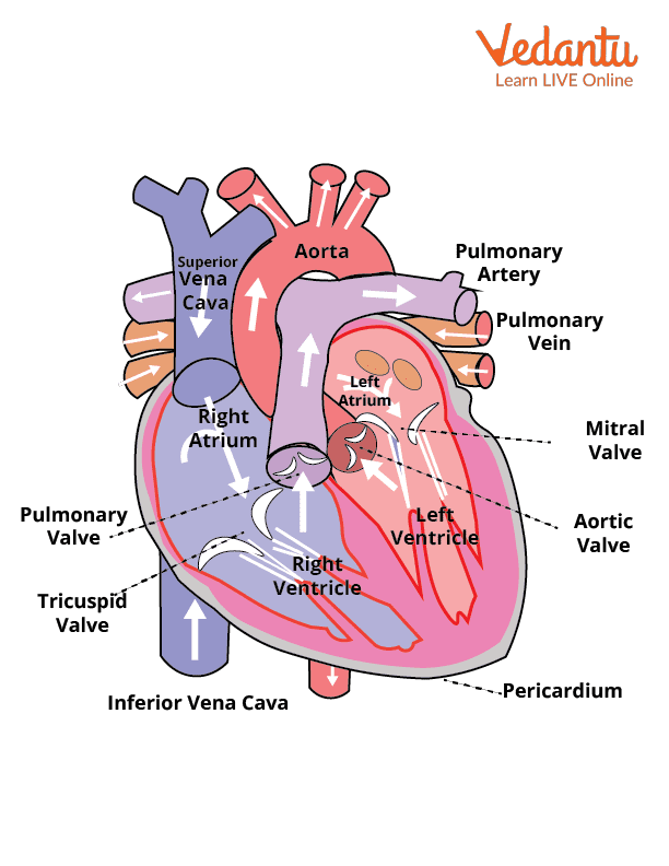
Heart Valves
The purpose of heart valves is to maintain a unidirectional blood flow through the heart. Instead of being constructed of muscle, the valves are formed of stiff connective tissue layers which function as flaps. Two AV valves (mitral valve and tricuspid valve) and two semilunar valves (aortic valve and pulmonary valve) make up the four-heart valve. Due to pressure variations along both sides of the valve, the valves gently open and close. The valve's leaflets are forced to open whenever there is increased pressure behind it, allowing the blood to flow inside. However, when the pressure in front of the valve is higher, the leaflets snap shut and blood flow is blocked.
Heart Valves
AV Valves
On each side of the heart, AV valves are located between the atrium and ventricles. These valves permit both the backflow of blood into the atrium during systole and the movement of blood from the atria to the ventricles. On the right side, the AV valve is named the tricuspid valve, which has three leaflets, while on the left side the valve is called as a mitral or bicuspid valve with two leaflets.
AV valves are forced to open and blood flows into the ventricles when the atrial pressure is higher than ventricular pressure. On the other hand, AV valves close rapidly when the ventricular pressure is greater than atrial pressure. The chordae tendineae, which are fibrous connective tissues which bind to the valves, prevent them from inverting.
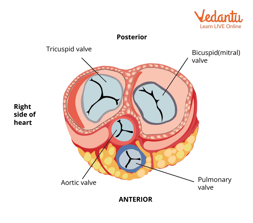
AV Valves
Semilunar Valves
Semilunar valves get their name from the fact that they resemble a half-moon. They are situated between two main arteries and the ventricles. The pulmonary valve is situated between the right ventricle and pulmonary artery, whereas the aortic valve is situated between the left ventricle and aorta. Semilunar valves open when the ventricular pressure rises above arterial pressure due to the ventricle’s contraction, and blood is pushed into the major arteries. On the other hand, Semilunar valves close when the ventricles relax because arterial pressure is higher than ventricular pressure. The AV valves vs semilunar valves is described in the table below:
What Is the Function of Valves?
The valves are open and close in response to the heart muscle contracting and relaxing. This permits alternating blood flow into the ventricles and atria, which helps in maintaining the proper blood flow and stops blood from flowing backwards.
Valve Disorders
Heart can suffer severe harm if the valves don't open and close properly, resulting in problems like the following:
Leaky Valve - The valve fails to close totally, allowing backward blood flow, so called regurgitation.
Stenosis - This entails restricted valve opening which doesn't open properly.
Atresia - It is a valve opening deformation that blocks the blood flow from the atria to the ventricle, or from the ventricle to the pulmonary artery or aorta.
Clinical Significance
The major purpose of heart valves is to ensure unrestricted, one-way blood circulation. Blood is pumped from the atria with low pressure to the ventricles with high pressure, which further supply the major arteries. During the constant movement of the cardiac cycle, compressive and longitudinal stresses cause valve tissue deformity, or dislocation. This deformity changes the sensitivity to physiological stimuli and susceptible genetics that can lead to valve failure.
Interesting Facts
It's interesting to note that every valve must be opened for blood to leave the chamber and closed to prevent blood from returning in the wrong direction. The valves may be harmed by disease occurrence or regular life span use.
Key Features
The major purpose of heart valves is to preserve unrestricted, one-way blood flow.
During the cardiac cycle, the AV and semilunar valves open and close, directing blood flow through the heart chambers and out to the rest of the body.
The differing framework of AV and Semilunar valves has a major influence on disorders and wellness.


FAQs on AV Valves and Semilunar Valves in the Human Heart
1. What are AV valves and semilunar valves?
The AV (atrioventricular) valves and semilunar valves are heart valves that regulate one-way blood flow through the heart. AV valves lie between the atria and ventricles, while semilunar valves are located at the exits of the ventricles.
- AV valves: Include the tricuspid valve (right side) and mitral/bicuspid valve (left side).
- Semilunar valves: Include the pulmonary valve and aortic valve.
- Both types prevent backflow of blood and maintain efficient circulation.
2. What is the function of AV valves in the heart?
The main function of the AV valves is to prevent the backflow of blood from the ventricles into the atria during ventricular contraction. They ensure one-way blood flow from atria to ventricles.
- Open during atrial systole to allow blood into ventricles.
- Close during ventricular systole to stop reverse flow.
- Supported by chordae tendineae and papillary muscles to prevent valve inversion.
3. What is the function of semilunar valves?
The semilunar valves prevent blood from flowing back into the ventricles after it has been pumped into the arteries. They ensure proper blood flow from the heart to the lungs and body.
- Pulmonary valve: Prevents backflow from the pulmonary artery into the right ventricle.
- Aortic valve: Prevents backflow from the aorta into the left ventricle.
- They close during ventricular diastole.
4. What is the difference between AV valves and semilunar valves?
The key difference between AV valves and semilunar valves is their location and structure in the heart. AV valves lie between atria and ventricles, while semilunar valves are located at the ventricular exits.
- Location: AV valves (atria–ventricle); Semilunar valves (ventricle–artery).
- Examples: Tricuspid and mitral vs. pulmonary and aortic.
- Structure: AV valves have chordae tendineae; semilunar valves do not.
5. How do AV valves open and close?
The AV valves open and close due to pressure changes between the atria and ventricles. They operate passively based on blood pressure differences.
- Open when atrial pressure is greater than ventricular pressure.
- Close when ventricular pressure rises during contraction.
- Chordae tendineae prevent the valve flaps from turning inside out.
6. How do semilunar valves work during the cardiac cycle?
The semilunar valves open and close in response to pressure changes during the cardiac cycle. They regulate blood flow from ventricles to arteries.
- Open during ventricular systole when ventricular pressure exceeds arterial pressure.
- Close during ventricular diastole when arterial pressure becomes higher.
- This prevents backflow into the ventricles.
7. What are the names of the AV valves?
The two AV valves are the tricuspid valve and the mitral (bicuspid) valve. They control blood flow between the atria and ventricles.
- Tricuspid valve: Located between right atrium and right ventricle.
- Mitral valve: Located between left atrium and left ventricle.
- The mitral valve has two cusps, while the tricuspid has three.
8. Why are semilunar valves called semilunar?
The semilunar valves are called semilunar because their cusps are shaped like half-moons. The term “semi” means half, and “lunar” refers to the moon.
- Each semilunar valve has three crescent-shaped cusps.
- The shape helps form a tight seal when the valve closes.
- This design efficiently prevents arterial backflow.
9. Do semilunar valves have chordae tendineae?
No, semilunar valves do not have chordae tendineae. Unlike AV valves, they function without tendinous cords.
- AV valves are supported by chordae tendineae and papillary muscles.
- Semilunar valves rely on their cup-shaped structure and arterial pressure.
- This structural difference reflects their different locations and functions.
10. What happens if AV or semilunar valves fail to function properly?
If AV valves or semilunar valves fail, blood may flow backward, causing a condition known as valvular regurgitation or valve stenosis. This disrupts normal circulation and reduces cardiac efficiency.
- Regurgitation: Valve does not close properly, leading to backflow.
- Stenosis: Valve becomes narrowed, restricting blood flow.
- Severe dysfunction can lead to heart enlargement or heart failure.


