

Difference between gram positive and gram negative bacteria structure and staining
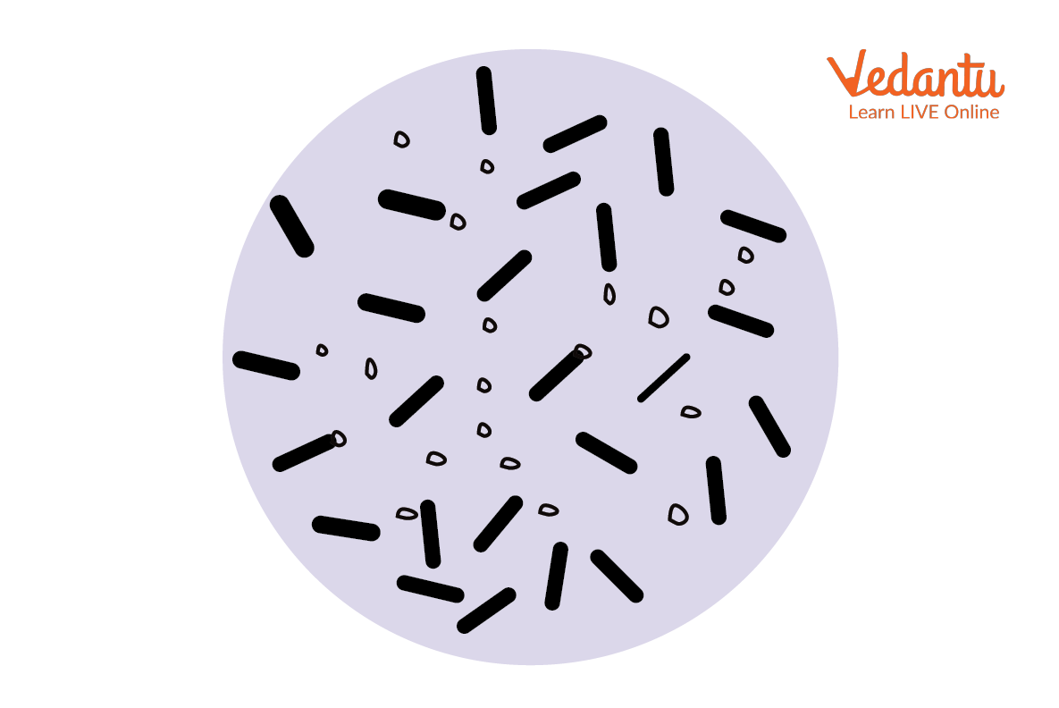
Bacteria are unicellular prokaryotic organisms devoid of nuclei. The size varies from 0.2 µm to 50 µm having varied morphology in their appearance. Examples of gram-positive bacteria are Methicillin-resistant Staphylococcus aureus (MRSA), and examples of gram-negative bacteria are Salmonella, Pneumonia and Gonorrhea. These groups of bacteria are a serious threat as they cause major diseases in humans.
All prokaryotic cells are surrounded by a complex cell made of peptidoglycan, also known as murein. In 1884, Christian Gram invented the staining procedure to classify bacteria. The bacteria that retain the Gram stain are gram-positive while which does not retain the gram stain are gram-negative bacteria. This difference lies in morphological variation seen in the cell wall structure of both gram-positive and gram-negative bacteria. Gram-positive and gram-negative bacteria difference pdf can be downloaded for more information.
Features of Gram-Positive Bacteria
The two major divisions of gram-positive bacteria are:
Phylum Actinobacteria - Have high GC content. Example: Streptomyces.
Phylum Firmicutes - Have low GC content. Examples: Bacillus and Clostridium.
In gram staining procedures, they appear purple yielding positive results.
Gram Stain Results in Gram-positive Bacteria
This group is composed of a thick, homogenous, singular, peptidoglycan layer (20 to 80 nm thick).
Gram-positive bacteria lack cellular appendages and are non-motile.
They generally appear round, rod or filamentous.
The general mode of reproduction is binary fission.
This group has the ability to form endospores.
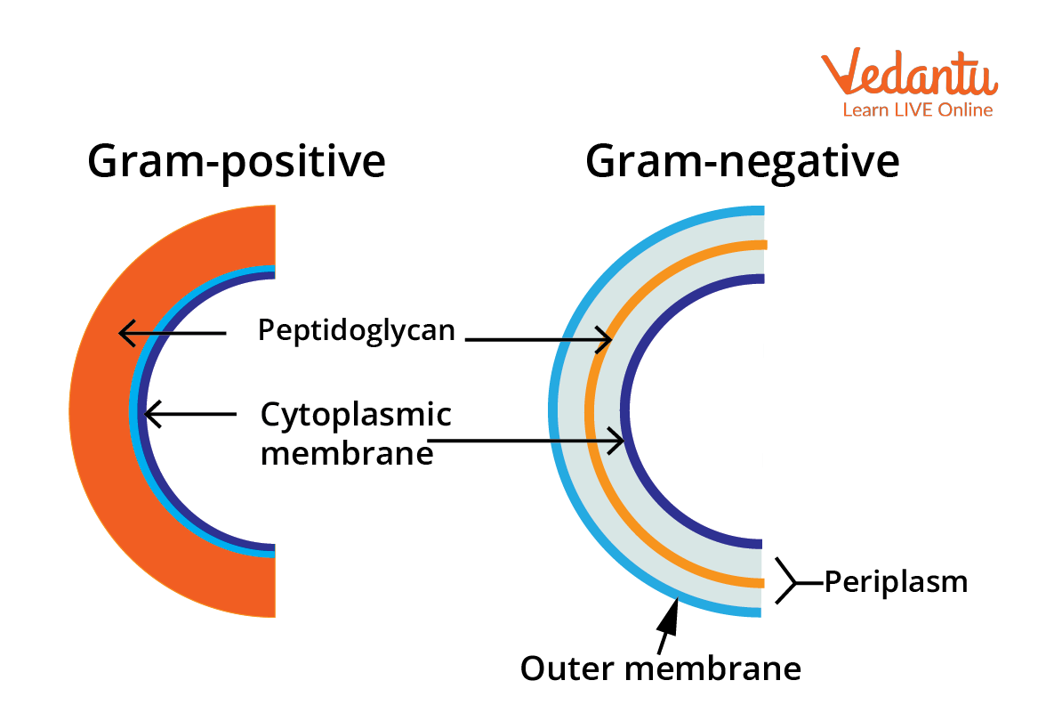
Cell Wall Structure of Gram-Positive Bacteria and Gram-Negative Bacteria
Features of Gram-Negative Bacteria
The gram-negative bacteria belong to the phylum Pseudomonadota. The two major groups are Enterobacteriaceae and the non-fermenters.
In gram staining, they appear red or pink as they do not retain the colour of crystal violet dye.
This group is composed of a 2 to 7 nm thick peptidoglycan layer surrounded by an outer membrane.
They are round, rod or spherical in shape.
Reproduction is either by binary fission or budding.
These bacteria do not form endospores.
Cell Wall Structure of Gram-Positive and Gram-Negative Bacteria
The cell wall of gram-positive and negative bacteria is made of peptidoglycan, also called murein. Peptidoglycan is a polymer having two sugar derivatives.
NAG - N-acetylglucosamine
NAM - N-acetylmuramic acid
These derivatives are linked together by β-1,4 glycosidic bonds. A carboxyl group of NAM tetrapeptide chains made of alternate D and L amino acids is linked. The peptide interbridge if present connects tetrapeptide chains. This peptide cross-link reaction is called transpeptidation.
The cell wall of gram-positive bacteria contains acidic negatively charged substances called teichoic acid. These are the polymers of glycerol and ribitol linked by phosphate moiety. The presence of teichoic acid makes the cell wall rigid by attracting positive ions like magnesium and sodium. In gram-negative bacteria, teichoic acid is absent.
Biosynthesis of Peptidoglycan
Linking of nucleotides with sugar precursors to form UDP-NAM and UDP-NAG; this step occurs in the cytoplasm.
Addition of amino acid group to UDP-NAM in a sequential manner to form NAM-pentapeptide.
In the third step, UDP-NAM pentapeptide forms a complex with bactoprenol to form lipid I. This step occurs at the plasma membrane.
A NAG molecule from a complex of UDP-NAG gets added to lipid I to form lipid II.
In the last stage, an outside plasma membrane polymerisation reaction occurs.
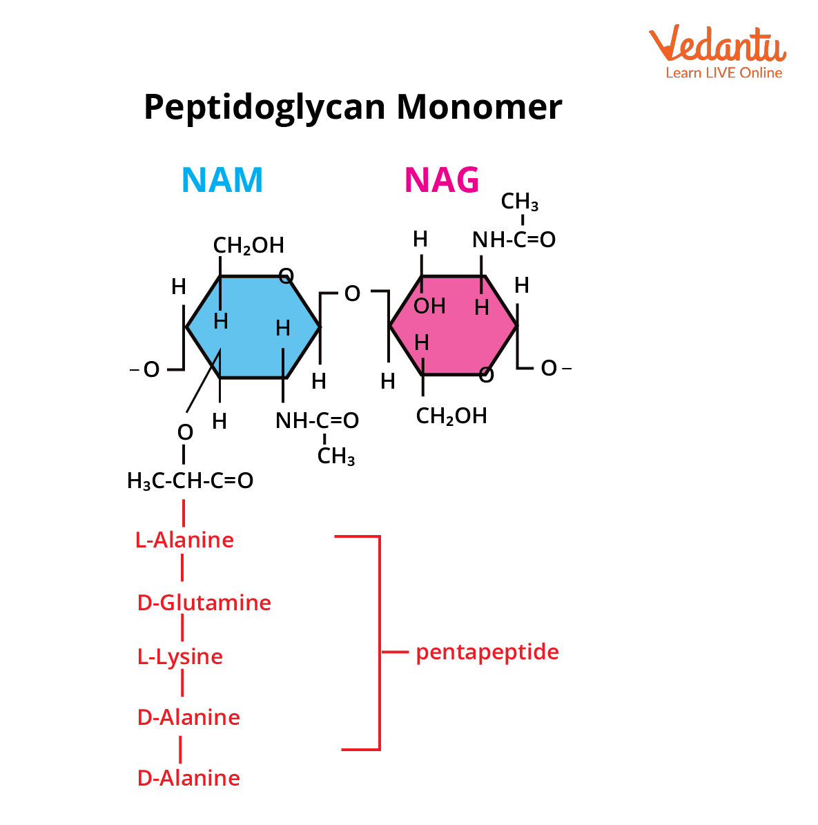
Peptidoglycan Monomer
Gram-negative bacteria have an additional outer membrane composed of lipids and polysaccharides. This complex is called the lipopolysaccharide complex (LPS). LPS acts as an endotoxin layer as it contains lipid A which is toxic. The outer membrane has porins which make it more permeable than the plasma membrane.
Differences between Gram-Positive and Gram-Negative Bacteria
Diagram of Gram-Positive and Gram-Negative Bacterial Cell Wall
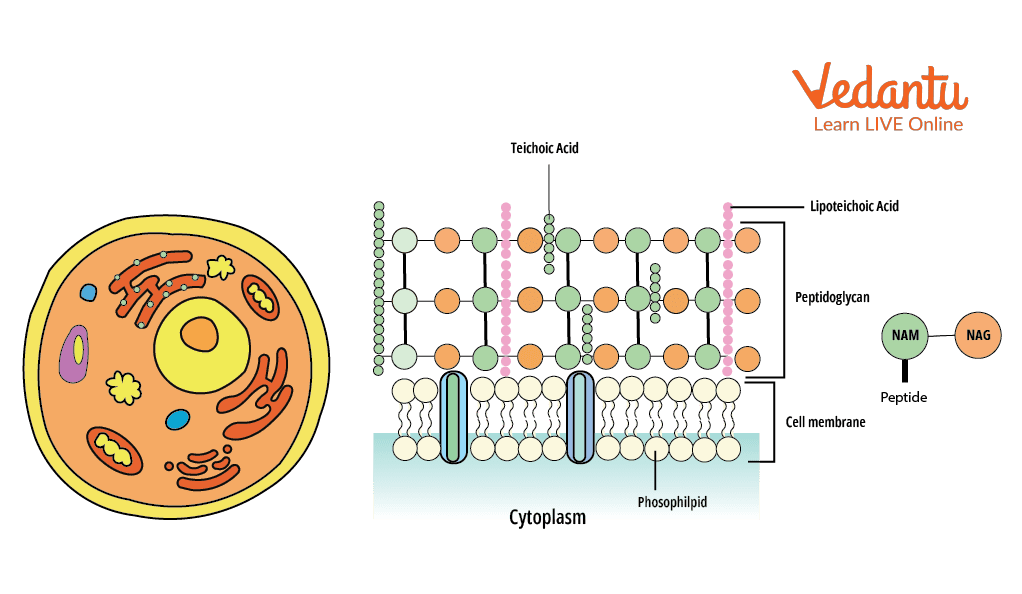
Structure of Gram-positive Cell Wall
Significance and Important Examples
Benefits and Examples of Gram-Positive Bacteria
These are non-pathogenic and are found in the human body.
Some of the gram-positive bacteria help in the formation of cheese.
Corynebacteria are used in the large-scale production of enzymes, nucleotides and amino acids.
Bacillus amyloliquefaciens acts as a natural antibiotic protein called barnase.
Negative Impact of Gram-Positive Bacteria
Staphylococcus species cause various skin-related disorders and are fomite borne. Similarly, gram-positive bacteria can cause food poisoning, respiratory disorders etc.
Negative Impact of Gram-Negative Bacteria
As these bacteria have outer membranes, they are resistant to the action of antibiotics. They act as endotoxin and cause diseases like cholera, meningitis, septic shock diseases etc.
Interesting Facts
According to WHO, a few pathogens, gram-positive bacteria are a serious concern and a health care problem, examples: multidrug-resistant (MDR) bacteria like methicillin-resistant Staphylococcus aureus (MRSA), vancomycin-resistant Enterococcus faecium (VRE) and β-lactamase-resistant Streptococcus pneumonia.
A new antibiotic named teixobactin produced by Eleftheria terrae was discovered in 2015. It is highly effective against resistant bacteria.
Important Questions
What are the differences between the cell walls of gram-positive and gram-negative bacteria?
Ans: The gram-positive bacteria have single-layered cell wall while gram-negative bacteria cell wall is multilayered having outer LPS membrane. This makes gram-negative bacteria more resistant.
What are the major diseases caused by gram-negative bacteria?
Ans: Diseases caused by gram-negative bacteria are a major concern to the healthcare industry as they are highly resistant to the action of antibiotics. Major infections include pneumonia, bloodstream infections, wound or surgical site infections, and meningitis.
Key Features
The cell wall of gram-positive and gram-negative bacteria is a chemical complex structure. It protects bacteria and plays a key role in adaptation.
The major component of the cell wall is peptidoglycan which is conserved in all prokaryotic organisms.
Gram-negative bacteria have outer membranes, thus, these groups of bacteria are human pathogens.


FAQs on Gram Positive and Gram Negative Bacteria Explained
1. What are Gram positive and Gram negative bacteria?
Gram-positive bacteria and Gram-negative bacteria are two major groups of bacteria classified based on their response to the Gram stain and differences in their cell wall structure.
- Gram-positive bacteria retain the crystal violet stain and appear purple due to a thick peptidoglycan layer.
- Gram-negative bacteria do not retain crystal violet and appear pink after counterstaining because they have a thin peptidoglycan layer and an outer membrane.
2. What is the difference between Gram positive and Gram negative bacteria?
The main difference between Gram-positive and Gram-negative bacteria lies in their cell wall structure and staining reaction.
- Gram-positive bacteria: Thick peptidoglycan layer, no outer membrane, stain purple.
- Gram-negative bacteria: Thin peptidoglycan layer, presence of outer membrane containing lipopolysaccharide (LPS), stain pink.
- Gram-negative bacteria are generally more resistant to antibiotics due to the protective outer membrane.
3. Why do Gram positive bacteria stain purple?
Gram-positive bacteria stain purple because their thick peptidoglycan layer traps the crystal violet–iodine complex during Gram staining.
- Crystal violet enters all bacterial cells.
- Iodine forms a complex with crystal violet.
- Alcohol decolorization shrinks the thick peptidoglycan layer.
- The dye complex remains trapped, so cells appear purple.
4. Why do Gram negative bacteria stain pink?
Gram-negative bacteria stain pink because the alcohol wash removes the crystal violet from their thin peptidoglycan layer, allowing the safranin counterstain to color them pink.
- The outer membrane is disrupted by alcohol.
- The thin peptidoglycan cannot retain the crystal violet–iodine complex.
- Safranin stains the decolorized cells pink.
5. What is the structure of the Gram positive cell wall?
The Gram-positive cell wall consists mainly of a thick peptidoglycan layer and teichoic acids.
- Multiple layers of peptidoglycan provide rigidity and shape.
- Teichoic acids help maintain cell wall stability and ion transport.
- No outer membrane is present.
6. What is the structure of the Gram negative cell wall?
The Gram-negative cell wall has a thin peptidoglycan layer located between the inner membrane and an outer membrane containing lipopolysaccharide (LPS).
- Thin peptidoglycan lies in the periplasmic space.
- The outer membrane contains LPS, which acts as an endotoxin.
- Porin proteins in the outer membrane regulate molecule entry.
7. What is the Gram staining process step by step?
The Gram staining process is a four-step differential staining method used to classify bacteria.
- Step 1: Apply crystal violet (primary stain).
- Step 2: Add iodine solution (mordant) to form a dye complex.
- Step 3: Wash with alcohol or acetone (decolorizer).
- Step 4: Apply safranin (counterstain).
8. Can you give examples of Gram positive and Gram negative bacteria?
Common examples of Gram-positive and Gram-negative bacteria differ in structure and disease association.
- Gram-positive bacteria: Staphylococcus aureus, Streptococcus pyogenes, Bacillus anthracis.
- Gram-negative bacteria: Escherichia coli, Salmonella typhi, Pseudomonas aeruginosa.
9. Why are Gram negative bacteria more resistant to antibiotics?
Gram-negative bacteria are more resistant to antibiotics because their outer membrane acts as a protective barrier.
- The outer membrane limits drug entry.
- Porin channels selectively allow small molecules.
- They may possess efflux pumps that expel antibiotics.
10. What is the importance of Gram staining in microbiology?
The importance of Gram staining lies in its ability to rapidly classify bacteria and guide diagnosis and treatment.
- Helps identify whether bacteria are Gram-positive or Gram-negative.
- Assists in selecting appropriate antibiotic therapy.
- Provides insight into bacterial cell wall structure and pathogenicity.


