

Key differences in cell wall structure staining mechanism and antibiotic response
Gram-positive and Gram-negative bacteria are the two main categories of bacteria. Scientists use a laboratory process called Gram staining to differentiate between these two groups. This distinction is based on the structure of their cell walls and their reaction to certain stains. Understanding this difference is crucial, as it affects how infections are identified and treated.
Difference Between Gram-Positive and Gram-Negative Bacteria
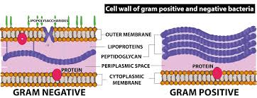
The main difference between Gram-positive and Gram-negative bacteria lies in the thickness of their cell walls and the presence of certain cell wall structures. Gram-positive bacteria possess a thick peptidoglycan cell wall, causing them to retain a blue or purple color after Gram staining. In contrast, Gram-negative bacteria have a thinner peptidoglycan wall and an additional outer membrane, resulting in a red or pink color after staining.
| Feature | Gram-Positive Bacteria | Gram-Negative Bacteria |
|---|---|---|
| Color after Gram staining | Blue or purple | Pink or red |
| Cell wall thickness | Thick peptidoglycan | Thin peptidoglycan, with outer membrane |
| Outer membrane | Absent | Present |
| Toxin type | Emetic, neurotoxin, enterotoxins | Endotoxins |
| Shapes | Spherical (cocci), rod (bacilli), or branching filaments | Sphere (cocci), rod (bacilli), spiral-shaped |
| Antibiotic resistance | Usually less resistant | Often more resistant |
| Common types |
Staphylococcus aureus, Staphylococcus epidermidis, Staphylococcus saprophyticus, Streptococcus pneumoniae, Streptococcus pyogenes, Enterococci, Corynebacterium diphtheriae, Bacillus anthracis |
Vibrio cholerae, Escherichia coli, Bartonella henselae, Campylobacter, Legionella, Salmonella, Salmonella typhi |
Understanding Gram Staining
Gram staining is a four-step process developed by Hans Christian Gram. It is used to classify bacteria and involves:
- Application of violet or methylene blue dye to bacterial cells.
- Cells are fixed and further stained, allowing the dye to enter their walls.
- Washing with a decolorizer (often alcohol).
- Application of a counterstain (typically red or pink).
Gram-positive bacteria retain the original dye and appear violet or blue. Gram-negative bacteria lose the dye during washing, then take up the counterstain and appear pink or red.
Characteristics and Health Significance
Gram-positive bacteria lack an outer membrane and are more likely to respond to antibiotics like penicillin. They may produce toxins such as emetic toxin or neurotoxins, leading to foodborne illnesses, skin infections, and other diseases.
Gram-negative bacteria possess an outer membrane that provides additional protection and resistance. Disruption of their wall can release endotoxins, sometimes causing severe reactions in the host. They are a leading concern in public health due to antibiotic resistance and their ability to cause outbreaks of diseases such as cholera and typhoid fever.
Examples of Gram-Positive Bacteria and Related Diseases
- Staphylococcus aureus – can cause skin infections and, in severe cases, blood infections.
- Staphylococcus epidermidis – major cause of catheter or device-related infections.
- Streptococcus pneumoniae – causes pneumonia and meningitis.
- Streptococcus pyogenes – responsible for throat infections, skin infections, and sometimes rheumatic fever.
- Enterococci – present in the intestines, but can lead to urinary tract or bile duct infections.
- Corynebacterium diphtheriae – causes diphtheria, affecting the throat and sometimes the skin.
- Bacillus anthracis – leads to anthrax, an infectious disease affecting both animals and humans.
Examples of Gram-Negative Bacteria and Related Diseases
- Vibrio cholerae – causes cholera, a serious diarrheal illness.
- Escherichia coli – can lead to diarrhea, urinary tract infections, or food poisoning.
- Bartonella henselae – causes cat scratch disease, transmitted by infected cats.
- Campylobacter – a major cause of bacterial diarrhea. Often contracted from undercooked poultry.
- Legionella – responsible for Legionnaires’ disease, a severe form of pneumonia acquired by inhaling contaminated water droplets.
- Salmonella (including Salmonella typhi) – causes food poisoning and typhoid fever.
Treatment and Antibiotic Resistance
Most Gram-positive infections are responsive to a broad range of antibiotics, including penicillin and tetracyclines. Gram-negative infections, because of the protective outer membrane, are often more challenging to treat. These bacteria may require more specific antibiotics, such as ciprofloxacin or azithromycin, and are known to become resistant to drugs more rapidly.
Preventing Antibiotic Resistance
Limiting antibiotic resistance starts with using antibiotics only when prescribed. People can help prevent resistance and the spread of infection by following hygiene practices, such as regular handwashing, food safety, keeping vaccinations up to date, and avoiding the use of leftover or unapproved antibiotics.
Summary Table
| Aspect | Gram-Positive | Gram-Negative |
|---|---|---|
| Stain color | Blue/purple | Red/pink |
| Wall structure | Thick peptidoglycan, no outer membrane | Thin peptidoglycan, with outer membrane |
| Resistance to antibiotics | Generally lower | Generally higher |
| Toxins | Produces exotoxins | Releases endotoxins |
Explore Related Biology Topics
Practice Question
Identify one key structural feature that makes Gram-negative bacteria more resistant to antibiotics than Gram-positive bacteria.
For more detailed explanations, examples, and interactive resources, explore biology lessons and practice exercises at Vedantu.


FAQs on Difference Between Gram Positive and Gram Negative Bacteria Explained
1. What is the difference between Gram positive and Gram negative bacteria?
The main difference between Gram positive bacteria and Gram negative bacteria is the structure of their cell wall and their response to Gram staining.
- Gram positive bacteria have a thick peptidoglycan layer and retain the crystal violet stain, appearing purple.
- Gram negative bacteria have a thin peptidoglycan layer and an additional outer membrane, appearing pink after staining.
- The outer membrane in Gram negative bacteria contains lipopolysaccharides (LPS), which can act as endotoxins.
2. What is Gram staining and how does it differentiate bacteria?
Gram staining is a differential staining technique used to classify bacteria based on differences in their cell wall composition.
- Step 1: Apply crystal violet (primary stain).
- Step 2: Add iodine (mordant) to form a dye complex.
- Step 3: Use alcohol or acetone for decolorization.
- Step 4: Apply safranin (counterstain).
Gram positive bacteria retain the violet color, while Gram negative bacteria lose it and take up the pink counterstain.
3. Why do Gram positive bacteria stain purple?
Gram positive bacteria stain purple because their thick peptidoglycan layer traps the crystal violet–iodine complex during decolorization.
- The thick cell wall prevents alcohol from washing out the stain.
- There is no outer membrane to disrupt dye retention.
- As a result, cells appear purple under a microscope.
4. Why do Gram negative bacteria stain pink?
Gram negative bacteria stain pink because the alcohol decolorizer removes the crystal violet from their thin peptidoglycan layer.
- The thin peptidoglycan cannot retain the dye complex.
- The outer membrane is disrupted by alcohol.
- They then absorb the safranin counterstain and appear pink or red.
5. What is the structure of the Gram positive bacterial cell wall?
The cell wall of Gram positive bacteria consists mainly of a thick peptidoglycan layer with embedded teichoic acids.
- Peptidoglycan provides rigidity and shape.
- Teichoic acids help in cell wall maintenance and ion transport.
- There is no outer membrane.
6. What is the structure of the Gram negative bacterial cell wall?
The Gram negative bacterial cell wall has a thin peptidoglycan layer and an additional outer membrane.
- The outer membrane contains lipopolysaccharides (LPS).
- A periplasmic space lies between the inner and outer membranes.
- The structure provides extra protection and antibiotic resistance.
7. What are examples of Gram positive and Gram negative bacteria?
Examples of Gram positive and Gram negative bacteria differ based on their cell wall structure and staining reaction.
- Gram positive bacteria: Staphylococcus aureus, Streptococcus pneumoniae, Bacillus subtilis.
- Gram negative bacteria: Escherichia coli, Salmonella typhi, Pseudomonas aeruginosa.
8. Why are Gram negative bacteria often more resistant to antibiotics?
Gram negative bacteria are often more resistant to antibiotics because their outer membrane acts as a protective barrier.
- The outer membrane limits drug entry.
- Porin proteins selectively allow molecules to pass.
- The periplasmic space may contain enzymes that degrade antibiotics.
9. What is the role of lipopolysaccharide (LPS) in Gram negative bacteria?
Lipopolysaccharide (LPS) is a major component of the outer membrane of Gram negative bacteria and acts as an endotoxin.
- It triggers strong immune responses in humans.
- The Lipid A portion is responsible for toxic effects.
- LPS also contributes to membrane stability and protection.
10. How is the difference between Gram positive and Gram negative bacteria important in medicine?
The difference between Gram positive and Gram negative bacteria is important in medicine because it guides diagnosis and antibiotic selection.
- Gram staining helps quickly identify bacterial type in infections.
- Certain antibiotics target peptidoglycan synthesis more effectively in Gram positive bacteria.
- Gram negative infections may require drugs that penetrate the outer membrane.


