

How Do Centrioles Help Cells Divide and Stay Organized?
Centrioles play a vital role in cell division and are typically found in pairs near the nucleus of many eukaryotic cells. They are best known for their function during mitosis and meiosis, where they help in organising spindle fibres. In this article, we will discuss what centrioles are, explore the characteristics of centrioles, understand their structure and functions, learn about their presence in plant cells, and address their significance in animal cells. We will also look at additional interesting facts and provide useful memory aids to help you learn more effectively.
What are Centrioles?
Centrioles are small, cylindrical cell organelles made up of microtubules. They are located near the nucleus and usually exist in pairs known as diplosomes. Although most animal cells contain two visible centrioles, certain lower plants such as Chlamydomonas also have them. By contrast, many higher plants (flowering plants) and conifers do not have centrioles at all.
Characteristics of Centrioles
They are cylindrical structures composed of microtubule triplets arranged in a circular pattern.
They normally occur in pairs and are oriented at right angles to each other.
They are central to the organisation of the microtubule organising centre (MTOC).
Cytokinesis and cell division processes are closely associated with centrioles in animal cells.
Despite not containing DNA, centrioles can duplicate themselves.
They are believed to help decide the spatial arrangement of the cell by dictating the position of the nucleus.
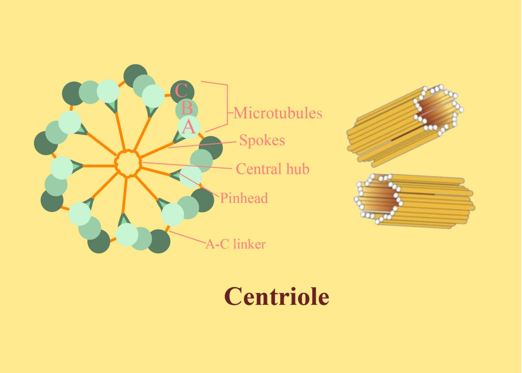
Centrioles Structure
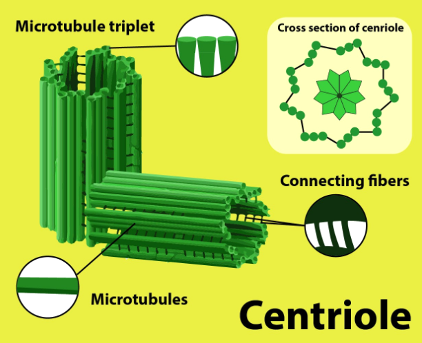
A typical centriole is made up of nine sets of microtubule triplets arranged in a cylindrical form. This arrangement is often referred to as the “9+0” pattern. Each triplet is composed of three microtubules tightly packed together.
Exceptions in Organisms
In the early embryo of Drosophila melanogaster, the centriole pattern may present 9 pairs of microtubules instead of triplets.
In C. elegans (roundworm), the sperm cell and early embryos may show nine single microtubules instead of triplets.
These structural details can only be observed under an electron microscope because the diameter of a centriole is very small.
Centrioles in Animal Cells
Centrioles are most commonly found in animal cells. They are crucial for forming spindle fibres during cell division (mitosis and meiosis). When an animal cell enters mitosis, each centriole pair migrates to opposite poles of the cell, contributing to the formation of spindle fibres that pull the chromosomes apart. This ensures the proper distribution of genetic material into the daughter cells.
Centrioles in Plant Cells
Most higher plant cells (angiosperms and conifers) lack centrioles. However, some lower plant forms like Chlamydomonas do possess centrioles. Instead of using centrioles, higher plants organise their spindle fibres using different structures such as polar organisers or nuclear envelope-associated microtubule organising centres. Despite the absence of centrioles in most plant cells, cell division proceeds normally with the help of microtubule networks.
Centrioles in Mitosis
During mitosis in animal cells, the centriole duplicates in the S phase of the cell cycle. As the cell nears division, the replicated pairs travel to opposite sides of the nucleus. Spindle fibres begin to form and attach to the chromosomes, separating them to ensure each daughter cell receives the correct set of genetic information. While it was once thought that centrioles were required for spindle formation, research indicates that cells (under experimental conditions) can sometimes form spindles without centrioles. However, having centrioles generally makes the process of spindle formation more efficient and organised.
Functions of Centrioles
Formation of Basal Bodies: Centrioles can transform into basal bodies, which act as anchoring sites for the growth of cilia and flagella in certain eukaryotes.
Cell Division: They participate in organising the spindle fibres essential for mitosis and meiosis. This helps in the accurate separation of chromosomes.
Determining Cell Polarity: The position of centrioles often determines the orientation of the nucleus and the overall architecture (polarity) of the cell.
Guiding Cytokinesis: After the chromosomes are separated, cytokinesis (the division of the cytoplasm) is coordinated with the help of microtubules associated with the centrioles.
Regulating Microtubule Organisation: They serve as the microtubule organising centre (MTOC), ensuring that microtubules are properly arranged in the cytoplasm.
Discovery of Centrioles
The presence of centrioles was observed in the late 19th century:
Edouard van Beneden (1883) and Theodor Boveri (1888) first identified centrioles in animal cells.
Later, in the 1950s, Joseph G. Gall and Etienne de Harven described how centrioles duplicate.
These discoveries were fundamental in understanding cell division mechanisms and the intricate organisation of microtubules in eukaryotic cells.
Additional Observations and Unique Facts
Removing centrioles by experimental methods (for example, through laser ablation) does not immediately stop cell division. However, the process may become less organised, indicating that centrioles greatly enhance the efficiency and stability of spindle formation.
In flagellated and ciliated organisms, the mother centriole directly gives rise to basal bodies that form the base of cilia or flagella.
Quick Quiz on Centrioles
Which of the following is the typical arrangement of microtubules in centrioles?
a) 9 pairs of microtubules
b) 9 sets of triplets of microtubules
c) 9 single microtubules
d) 8 sets of triplets of microtubules
Answer: b) 9 sets of triplets of microtubules
In which phase of the cell cycle do centrioles usually replicate?
a) G1 phase
b) S phase
c) G2 phase
d) M phase
Answer: b) S phase
Which organelle do centrioles transform into for the formation of cilia and flagella?
a) Mitochondria
b) Nucleus
c) Basal bodies
d) Golgi bodies
Answer: c) Basal bodies
Which of these organisms typically lacks centrioles?
a) Chlamydomonas
b) Many angiosperms
c) Animal cells
d) Fungi that produce zoospores
Answer: b) Many angiosperms
What is the main function of centrioles during mitosis?
a) Protecting the nucleus
b) Forming spindle fibres
c) Producing ATP
d) Storing enzymes
Answer: b) Forming spindle fibres


FAQs on Centrioles: Key Functions and Importance in Cells
1. What exactly are centrioles and where are they located inside a cell?
Centrioles are small, cylindrical organelles typically found in pairs within the cytoplasm of most animal cells and some lower plant cells. They are located near the nucleus in a specialised region called the centrosome, which acts as the main microtubule-organising centre of the cell.
2. What is the basic structure of a centriole?
A centriole has a unique structure often described as a 'cartwheel' or a '9+0' arrangement. This means it is made of nine sets of triplet microtubules arranged in a circle, with no microtubules in the centre. These microtubules are composed of a protein called tubulin.
3. What are the main functions of centrioles in a cell?
Centrioles play a few critical roles in the cell. Their primary functions include:
- Organising the microtubules that form the cell's cytoskeleton.
- Forming the spindle fibres required to separate chromosomes during cell division (mitosis and meiosis).
- Acting as basal bodies that anchor and form cilia and flagella, which help in cell movement.
4. Why are centrioles considered so important for cell division in animals?
During cell division, centrioles replicate and move to opposite poles of the cell. They organise the formation of the mitotic spindle, a network of fibres that attaches to chromosomes. These fibres then pull the chromosomes apart, ensuring that each new daughter cell receives a complete and correct set of genetic material.
5. What is the difference between a centriole and a centrosome?
This is a common point of confusion. A centrosome is the entire organelle that organises microtubules. It consists of two centrioles arranged perpendicular to each other, surrounded by a mass of protein called the pericentriolar material. So, centrioles are the core components within a centrosome.
6. If centrioles are so important for division, how do plant cells manage without them?
Higher plant cells and fungi lack centrioles but still divide successfully. They use other structures, broadly known as microtubule-organising centres (MTOCs), to form the spindle fibres. These centres are not as focused as a centrosome but effectively perform the same function of organising chromosomes for division.
7. How do centrioles help in the formation of structures like cilia and flagella?
Centrioles can migrate to the cell membrane and function as basal bodies. A basal body acts as a foundation or anchor from which a cilium or flagellum grows. The microtubule arrangement of the centriole provides the structural template for the growing cilium or flagellum.
8. What could happen if a cell's centrioles were damaged or absent?
If centrioles are damaged, the formation of the spindle during cell division can be disorganised. This can lead to serious errors, such as an incorrect number of chromosomes in the daughter cells (aneuploidy), which may cause the cells to die or become cancerous. It could also affect the cell's ability to form cilia or flagella.


