


Class 12 Biology Chapter 2 Summary Notes PDF Download
Human Reproduction Class 12 Notes simplify learning for students. These notes thoroughly cover essential topics like gametogenesis, the menstrual cycle, fertilisation, and embryonic development. Complex concepts are broken down into easy-to-understand explanations, summaries, and key facts. Diagrams and examples are provided to enhance clarity. Class 12 Biology Notes are ideal for quick revision and exam preparation, helping students grasp the fundamentals and excel in their exams.
 Table of Content
Download the FREE Human Reproduction Class 12 Short Notes PDF from Vedantu, updated according to the latest CBSE Class 12 Biology Syllabus, for effective study sessions.


Section–A (1 Mark Questions)
1. Where are seminiferous tubules located in human body?
Ans. Seminiferous tubules are located within the testes. These are the sites for production of male gametes, namely spermatozoa.
2. Which glands secrete the primary male sex hormone?
Ans. Testes have a distinction of being an endocrine gland also because they secrete testosterone, the primary male sex hormone that is required for the normal development of male physical characteristics.
3.Write the names of following.
a. Site for maturation of sperms
b. Embryo at 16 cell stage
Ans. a. Seminal vesicles b. Morula
4. What is spermiogenesis?
Ans. Spermiogenesis is formation of spermatozoa from spermatids. It is the one of stages of spermatogenesis. The spermatids then transformed into spermatozoa.
5. What induces foetal ejection reflex in humans?
Ans. Fully developed foetus and placenta induce parturition signals, which leads to mild uterine contractions and hence foetal ejection reflex.
Section–B (2 Marks Questions)
6. If a primary spermatocyte undergoes first meiotic division to form two secondary spermatocytes than how many secondary spermatocytes are required to form 400 spermatozoa? Explain with reason.
Ans. A primary spermatocyte undergoes first meiotic division (reductional division) to form two equal, haploid cells called secondary spermatocytes. Each secondary spermatocyte undergoes the second meiotic division (equational division) to produce two equal, haploid spermatids. Each spermatid is transformed into one spermatozoon (sperm). Since each secondary spermatocyte gives 2 spermatozoon, 200 secondary spermatocytes are required to produce 400 spermatozoa.
7. Testicular lobules in each testis contain highly coiled seminiferous tubules. Answer the following questions regarding seminiferous tubules.
(i) What are the two types of cells present on the inner lining of seminiferous tubules?
(ii) What are the functions of these cells?
Ans. (i) The inner side of seminiferous tubules are lined with epithelium, and it contains two types of cells: the male germ cells (spermatogonia) and Sertoli cells.
(ii) The spermatogonia undergo meiotic divisions which leads to formation of sperms (the male gametes). Sertoli cells are present in between the germinal epithelial cells of the seminiferous tubules and these cells provide nourishment to the developing spermatozoa (sperms)
8. What is colostrum and why is it essential for new-born babies?
Ans. Colostrum is sticky yellow fluid which is secreted in the breastmilk during the few days after birth of the baby. It contains several antibodies that are essential for development of innate immunity in the foetus. Colostrum is also rich in proteins and has a low-fat content, which is required for development of the new-born babies.
9. Name the locations and functions of the following
(i) Corpus luteum
(ii) Endometrium
Ans. (i) Corpus luteum: The corpus luteum is a yellow-coloured glandular body which is formed from the remnants of the graafian follicle after ovulation. It is located within the ovary and secretes progesterone which helps to maintain the endometrial layer in the uterus. Corpus luteum degenerates in the absence of fertilization.
(ii) Endometrium: It is the inner glandular layer that lines the uterine cavity. It undergoes cyclical changes during the menstrual cycle. This layer helps in implantation of fertilized ovum. It sheds with unfertilized ovum during menstruation.
10. What happens in the follicular phase of the menstrual cycle?
Ans. Follicular phase is also known as proliferative phase. In this phase the primary follicles in the ovary become a mature Graafian follicle and simultaneously in the uterus the endometrium regenerates through proliferation. These changes are induced by changes in the levels of pituitary and ovarian hormones. The secretion of gonadotropins (LH and FSH) increases gradually during the follicular phase and stimulates follicular development as well as secretion of oestrogen by the growing follicles.
11. What are the main functions of testis and ovary?
Ans. Testes also called as the testicles are two oval-shaped organs in the male reproductive system. Testes are responsible for the production of sperms and the male hormone testosterone. They also serve as storage organs of sperms until they’re mature enough for ejaculation.
The ovaries are part of the female reproductive system. Ovaries produce and store the ovum or the egg cell. They also produce the female sex hormone oestrogen.
PDF Summary - Class 12 Biology Human Reproduction Notes (Chapter 2)
Basic Steps in Human Reproduction:
1. Gametogenesis
2. Insemination
3. Fertilization
4. Blastocyst development
5. Implantation
6. Embryo development
7. Parturition
2.1 Male Reproductive System
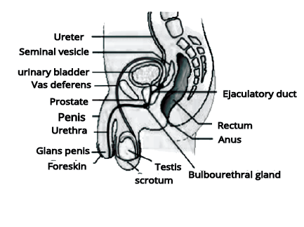
There are four main parts in the male reproductive system:
1. Testes
2. Accessory ducts
3. Glands
4. External Genitalia
Testes (Singular Testis):
Situated in the pelvic region outside the abdominal cavity within a pouch called a scrotum.
A small muscular sac is known as Scrotum that contains and protects the testes. The scrotum is located behind the penis and is considered a part of the external male genitalia.
The testes are sited outside the abdominal cavity to maintaining the low temperature of the testes nearly 2-2.5◦C lower than the average human body temperature. This condition is necessary for the synthesis of sperms.
The lower temperature in testes is required for spermatogenesis as the normal human body temperature can lead to mutation in the sperms.
Testis is oval in shape with the length of 4-5cm and 2-3cm wide.
The testis contains about 250 compartments known as Testicular lobules.
Each lobule normally comprises 1-3 highly coiled seminiferous tubules that play a significant role in sperm production.
Seminiferous tubules are the place that forms spermatozoa by the process of meiosis.
Each seminiferous tubule contains two types of cells i.e., spermatogonia and Sertoli cells in their inner lining.
These spermatogonia are diploid in nature and called the immature germ cells and they form the sperm by the process of meiosis. They contain 46 chromosomes in their cells.
Sertoli cells in seminiferous tubules provide nutrition to the spermatogonia.
Interstitial spaces are the region that presents outside the seminiferous tubule that contains the small blood vessels, Leydig cells, and some immunocompetent cells.
Leydig cells synthesize and secrete the testicular hormones called androgens.
Accessory Ducts:
The accessory ducts facilitate the transportation of the sperms from the testes to the urethra for their release outside the body. The male reproductive system consists of four accessory ducts:
Rete Testis
Vasa efferentia
Epididymis
Vas deferens
Rete Testis: These are the ducts where the seminiferous tubules open into a series of channels.
Vasa Efferentia: These ducts make a pathway to transport the sperm from the rete testis to the epididymis which is placed in the posterior surface of each testis.
Epididymis: It is a long, coiled tube that connects a testicle to a vas deferens. The Epididymis is sited on the backside of each testicle.
Vas Deferens: Vas deferens is a muscular duct that ascends into the abdominal cavity and makes a loop over the urinary bladder. It transports the sperm from the epididymis to the ejaculatory duct.
Ejaculatory Duct: This duct is situated on each side of the prostate gland. Ejaculatory ducts store and transport the sperms from the testis to the outside via the urethra.
Urethra: Urethra is a thin muscular tube that originates from the urinary bladder and then passes through the penis to its external opening known as the urethral meatus.
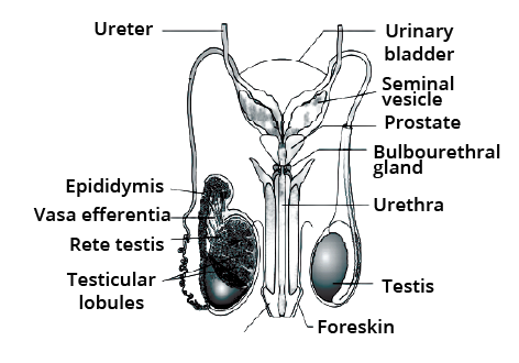
Figure: Male reproductive system
Accessory Glands
There are mainly three types of accessory glands:
Seminal vesicles
Prostate gland
Bulbourethral glands
These glands secrete the seminal plasma that mainly contains fructose, calcium, and certain enzymes. This secretion mixes with the sperms to nourish and protect them.
Seminal Vesicles: The Seminal vesicles contribute a significant proportion of approximately 60-75% of the fluid in semen. This fluid is rich in enzymes, proteins, vitamin C, fructose, prostaglandins, and phosphorylcholine. The high fructose content gives nutrient energy to the spermatozoa.
Prostate Gland: The prostate gland is a dense structure that is placed just inferior to the urinary bladder. The secretion of the prostate gland is slightly alkaline, thin, and milky colored. It helps in the survival of sperms in the acidic vaginal environment and also improves the motility of the sperms.
Bulbourethral Glands: The secretion of these glands helps in the lubrication of the penis and also neutralizes any residual acidity in the urethra.
2.2 Female Reproductive System:
The human female reproductive system is specialized to carry out various functions like gametogenesis, ovulation, fertilization, pregnancy, birth, and child care. The female reproductive system is made up of several parts:
1. A pair of ovaries
2. A pair of oviducts
3. Uterus
4. Cervix
5. Vagina
6. External genitalia
7. Mammary glands
Ovaries:
The ovaries are small and oval-shaped and are known as primary female sex organs.
They produce the female gamete generally called ovum and also produce ovarian hormones.
The ovaries are about 2-4cm in length.
They are connected to the pelvic wall and uterus with the help of ligaments.
The ovaries are protected by a thin epithelium which encircles the ovarian stroma.
Ovarian stroma- It is the matrix of the ovary which is divided into two regions:
1. Peripheral cortex
2. Inner medulla
3. Accessory Ducts:
The accessory ducts of the female reproductive system include the oviducts, uterus, and vagina.
Oviduct (Fallopian Tube):
A fallopian tube is generally 10-12 cm in length.
It covers from the periphery region of each ovary to the uterus.
Infundibulum: It is a proximal part of the oviduct that is present closer to the ovary. It is funnel-shaped and possesses fimbriae.
Fimbriae: They are the finger-like projections located at the edges of the infundibulum. They help in the collection of the ovum after ovulation
Ampulla: This is the wider part of the oviduct that connects with the infundibulum.
Isthmus: This is the last part of the oviduct that extends through the uterine walls and opens into a narrow lumen.
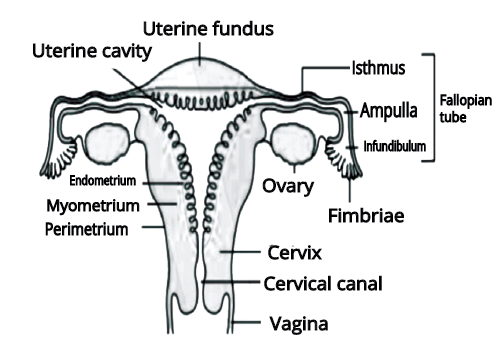
Figure: Female reproductive system
Uterus:
The uterus has also been named the womb.
It is designed like an inverted pear and attached to the pelvic wall by ligaments.
In the uterus, the embryo develops into the foetus.
It opens into the vagina via a narrow cervix.
Uterine wall possesses three layers of tissues- perimetrium, myometrium, and the endometrium
Perimetrium: This is the thin external membranous layer that protects the uterus from friction.
Myometrium: It is the thick middle layer that is made up of smooth muscle. It shows strong contractions at the time of the delivery of the baby
Endometrium: This is the inner glandular layer that lines the uterine cavity. It undergoes periodic changes during the menstrual cycle.
Cervix:
It is a narrow canal usually 2 to 3 cm in length that connects the uterus to the vagina.
Cervical Canal: The cervical canal connects the cavity of the body of the uterus with the lumen of the vagina.
Birth Canal: The cervical canal along with the vagina forms the birth canal which helps during the birth of the baby.
External Female Genitalia:
The main external female genitalia of the female reproductive system consists of the mons pubis, labia majora, labia minora, hymen, and clitoris.
Mons Pubis: It is a cushion of fatty tissue which is covered by hair and skin.
Labia Majora: They are fleshy folds of tissue that enclose and protect the external reproductive organs. It extends from the mons pubis and covers the vaginal opening.
Labia Minora: They are the paired folds of tissue that lies just inside the labia majora
Hymen: Hymen is a thin membrane that surrounds the opening of the vagina. It is often torn during the first intercourse or coitus or it can also be broken by participation in some sports like horseback riding, cycling, apart from this a sudden fall or jolt, insertion of a vaginal tampon is also the reason for the broken hymen. In some women, the hymen can continue even after coitus so it shows that the presence or absence of a hymen is not a reliable indication of virginity or sexual experience.
Clitoris: It is a small, sensitive, finger-like projection that is present at the junction of the labia minora above the urethral opening.
Mammary Glands:
All-female mammals possess a pair of functional mammary glands. The breasts are paired structures that are made up of glandular tissue and variable amounts of fats. Each mammary gland consists of 15-20 mammary lobes. These mammary lobes contain clusters of cells known as alveoli. The secretion of milk takes place by the cells of the alveoli. This secreted milk is stored in the lumen or cavities of the alveoli which open into the mammary tubules. These mammary tubules of each lobe join to form a mammary duct.
2.3: Gametogenesis:
It is the process by which the primary male and female sex organs like the testes in males and the ovaries in the female produce gametes.
Spermatogenesis: This is the process by which the immature male germ cells also known as spermatogonia produce mature sperm cells in the testis.
Oogenesis: Oogenesis is the process by which the immature oogonia in the ovaries produces a mature ovum.
Spermatogenesis: The spermatogenesis process starts at puberty and proceeds as follows:
First of all, the spermatogonia multiply by mitosis to increase in number. They are present inside the inner wall of seminiferous tubules where they multiply by mitotic division. Each spermatogonium is diploid in nature and contains 46 chromosomes.
Some spermatogonia known as the primary spermatocytes occasionally undergo meiosis to form two equal, haploid cells. These cells are called secondary spermatocytes. Due to their haploid nature, they contain 23 chromosomes.
The secondary spermatocytes further undergo second meiotic division and produce four haploid spermatids that also contain 23 chromosomes.
The spermatids then undergo spermiogenesis to produce sperms (spermatozoa). The heads of the sperms are embedded in the Sertoli cells.
The sperms are finally free from the seminiferous tubules by the process of spermiation.
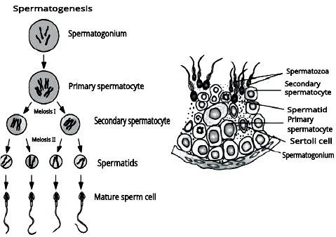
Spermatogonia: They are the immature male germ cells that undergo meiotic divisions to form sperms. Spermatogonium (singular) is diploid in nature and contains 46 chromosomes.
Primary Spermatocytes: These are the spermatogonia that undergo the meiosis process to form two equal haploid cells called secondary spermatocytes. They contain 46 chromosomes in each cell.
Secondary Spermatocytes: They are haploid in nature and arise from the primary spermatocytes as a result of meiosis I. They comprise 23 chromosomes.
Spermatids: These cells are arising from the secondary spermatocytes as a result of meiosis II. Spermatids are haploid cells that contain 23 chromosomes.
Spermiogenesis: It is the process by which spermatids mature to form spermatozoa or sperms.
Spermiation: This is the process by which sperms are released from the seminiferous tubules.
Hormones Affecting Spermatogenesis:
Gonadotropin-releasing hormone (GnRH): Gonadotropin-releasing hormone is the hypothalamic factor. The levels of this hormone increase mainly at puberty. The secretion of GnRH stimulates the release of two gonadotropins from the anterior pituitary – Follicle Stimulating Hormone (FSH) and Luteinizing Hormone (LH).
Follicle Stimulating Hormone (FSH): This hormone acts on the Sertoli cells where it stimulates the release of some factors that play an important role in the process of spermatogenesis.
Luteinizing Hormone (LH): LH is important in regulating the function of testes and ovaries. It acts on the Leydig cells where it stimulates the synthesis and secretion of androgens.
Structure of Sperm:
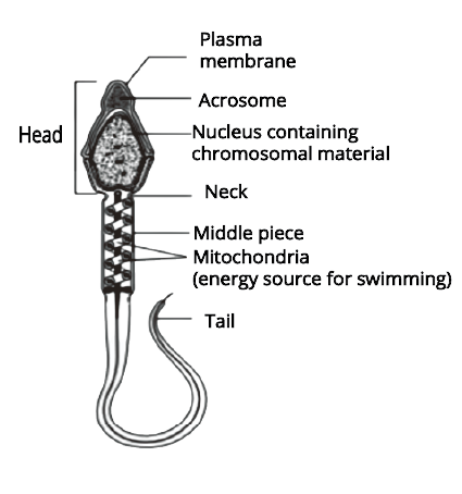
Sperm is a male reproductive cell that is made up of a head, neck, middle piece, and tail.
The whole body of the sperm is covered by a plasma membrane
Head: The head portion of the sperm contains an elongated, haploid nucleus
The anterior portion of the head contains an acrosome which contains some enzymes that help in dissolving the membrane of the egg cell and help in fertilization of the ovum.
Middle Piece: It comprises several mitochondria that provide energy for the vigorous motility of the tail. The movement of the tail is essential for fertilization because it helps sperm to move towards the ovum.
Approximately 200-300 million sperms are released during ejaculation. Out of these, at least 60% of them should have normal size and shape and at least 40% should show vigorous motility.
Semen: It is a milky white organic liquid that is released by the penis at the time of ejaculation. It includes sperms and the fluids secreted by the accessory ducts and the accessory glands like epididymis, vas deferens, prostate, seminal vesicles, and the bulbourethral glands.
The testicular hormones also known as androgens maintain the functions of the male accessory ducts and glands.
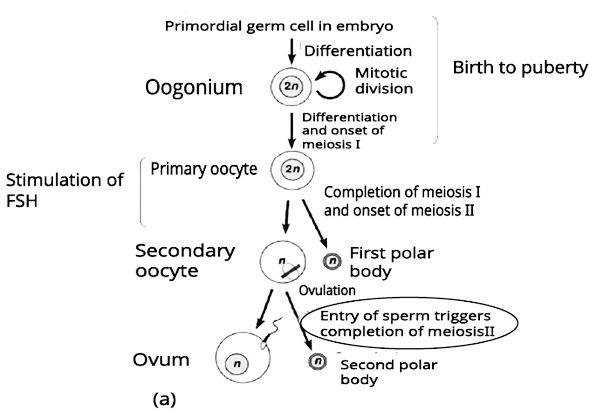
Oogenesis:
The process of formation of a mature female gamete is known as Oogenesis. It is introduced during the embryonic development stage. At this stage, a few million gamete mother cells or oogonia are formed in the fetal ovary. After birth, no more oogonia are formed or added.
The oogonia form primary oocytes that form by the process of meiosis and get arrested at the stage of Prophase.
Each primary follicle is formed by primary oocytes which are surrounded by a layer of granulosa cells.
Between birth to puberty, a large number of primary follicles get degenerated. Therefore, there are only about 60,000-80,000 primary follicles available in the ovary at the time of puberty.
These remaining primary follicles get surrounded by a few more layers of granulosa cells as well as a new theca to form the secondary follicle.
The tertiary follicle is formed by the secondary follicle. It is characterized by the presence of a fluid-filled space called an antrum and two layers of the theca. The layers of theca are arranged into two layers-inner theca interna and the outer theca externa. The primary oocyte increases in size and completes its first meiotic division. This division is an unequal division that forms a large secondary oocyte and a tiny first polar body.
Secondary oocyte consists of much of the nutrient-rich cytoplasm and it also develops a thick covering known as zona pellucida.
The tertiary follicle is finally converted into the mature Graafian follicle.
This Graafian follicle now ruptures and releases the secondary oocyte or ovum from the ovary. The process of release of the ovum is called ovulation.
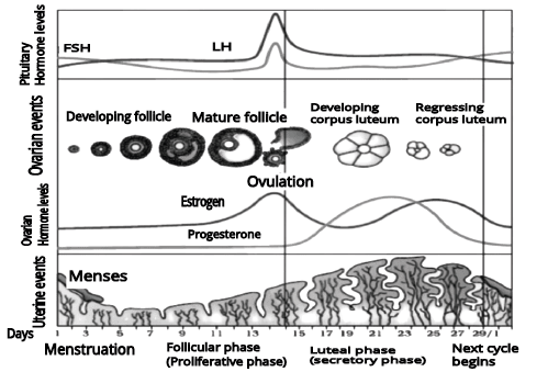
2.4 Menstrual Cycle:
Menstrual Cycle: This is the reproductive cycle that starts from one menstruation till the next one. It mainly occurs in female primates like monkeys, apes, and human beings. The cycle repeats at an interval of 28-35 days and normally releases one egg per cycle. This cycle is important for the production of oocytes and for the preparation of the uterus for pregnancy.
Menstruation: In this process, the blood and mucosal tissue are regularly discharged in a periodic manner. It occurs due to the breakage of the inner lining of the uterus. This process takes place once a month and is called a period.
Menarche: Menarche is the first menstruation for a human female that begins at puberty. The actual age for menarche generally differs from person to person. The first menstruation is the signal of the beginning of reproductive age in females.
Menopause: Menopause is defined as the permanent ceasing of the menstrual cycle in females. It occurs due to the depletion of oocytes and the loss of the ability of the ovary to produce estrogen as a result of aging. menopause. The average age of menopause is between 45-50 years and it varies from person to person.
Phases of the Menstrual Cycle:
The menstrual cycle follows four phases:
1. Menstrual phase
2. Follicular phase
3. Ovulation
4. Luteal Phase
1. Menstrual Phase: In this phase, the menstrual flow takes place.
The flow naturally lasts for 3-5 days.
In this phase, the breakdown of the endometrial lining of the uterus along with blood vessels forms a red liquid substance that comes out of the vagina.
This process only takes place in the condition when the ovum released by the ovary is not fertilized.
At the time of pregnancy, the menstrual flow does not occur.
Some factors like stress, poor diet, poor health, etc. affect menstruation.
2. Follicular Phase: In this phase, maturation of Graafian follicle occurs.
In the ovary, the primary follicles grow to become a fully mature Graafian follicle.
The endometrium lining of the uterus regenerates by proliferation.
These changes in endometrium and follicular regeneration are induced by ovarian hormones- Luteinizing Hormone (LH) and the Follicle Stimulating Hormone (FSH).
The secretion of LH and FSH gonadotropins increases gradually in this phase to stimulate follicular development. The secretion of estrogens by the growing follicles is also stimulated by this increased level of LH and FSH.
3. Ovulation/Ovulatory Phase: This is the phase where the release of the ovum from the ovary takes place.
In this phase, the levels of LH and FSH attain a peak at mid-cycle, around the 14th day.
LH Surge: The rapid secretion of LH leading to maximum LH level in mid cycle is called the LH surge. It induces the rupture of the Graafian follicle to release the ovum.
Ovulation: The release of an ovum from a mature Graafian follicle is known as ovulation. It occurs due to the LH surge which takes place in mid-cycle around the 14th day.
4. Luteal Phase: This is the phase of formation of the corpus luteum and it is also known as the secretary phase.
The ruptured parts of the Graafian follicle after ovulation form the yellow body called corpus luteum.
It secretes large amounts of progesterone hormone which maintains the endometrium. The endometrium is required for the implantation of the fertilized ovum and other events of pregnancy.
During pregnancy, there is no menstruation because of all events of the menstrual cycle stop.
In absence of fertilization, the corpus luteum degenerates. This causes the disintegration of the endometrium leading to menstruation and hence a new cycle starts.
2.5 Fertilisation and Implantation:
Insemination: It is the process in which semen is released into the vagina by the penis during coitus or copulation.
The motile male gamete or sperm swims rapidly through the cervix, enters into the uterus, and finally reaches the site of fertilization which is in the ampullary region of the fallopian tube.
The ovum which is released by the ovary is also transferred to the ampullary region of the fallopian tube.
Fertilization takes place in the fallopian tube only when the ovum and sperms are simultaneously transferred into the site of fertilization.
Fertilization: It is the fusion of the haploid male gamete or sperm and the haploid female gamete or egg. At the time of fertilization, the sperm makes changes in the zona pellucida layer of the ovum to block the entry of other sperms. This ensures that only one sperm can fertilize a single ovum.
The secretions of the acrosome make easy the entry of the sperm to the ovum by the zona pellucida and the plasma membrane.
This phenomenon induces the secondary oocyte to complete meiosis. This is again an unequal division and forms a second polar body and a haploid ovum.
The haploid nucleus of the sperm and the ovum fuse together and form a diploid zygote. Hence, a zygote contains 46 chromosomes.
The sex chromosome in the sperm is the factor that determined the sex of the fetus. As the female is XX the ovum will always carry the X chromosome, however, Males are XY and therefore, the sperm can contain either X or Y. Hence, half of all the sperms carry the X chromosome, and the remaining half carry the Y chromosome.
The zygote that carries an XY would develop into a male while XX would develop into a female.
The zygote is divided by the mitotic division which starts as it moves along the isthmus of the oviduct towards the uterus and forms 2, 4, 8, and 16 daughter cells known as blastomeres.
Morula: The embryo with 8-16 blastomeres is known as a morula.
It continues its division as it moves further along into the uterus. The blastomeres are arranged into two layers. First is the outer layer called the trophoblast and the other is the inner cell mass which is attached to the trophoblast.
The trophoblast layer then attaches to the endometrium of the uterus.
The inner cell mass differentiates to cover the blastocyst.
The cells of the blastocyst embedded in the uterine wall. This phenomenon is called implantation which leads to pregnancy.
Pregnancy and Embryonic Development
Chorionic Villi and Placenta:
After implantation, chorionic villi form on the trophoblast, surrounded by uterine tissue and maternal blood.
The chorionic villi and uterine tissue interlock to form the placenta, which connects the developing embryo to the mother.
The placenta provides oxygen and nutrients to the embryo and removes waste products.
Umbilical Cord:
The placenta is connected to the embryo by the umbilical cord, which transports substances to and from the embryo.
Placenta as Endocrine Tissue:
The placenta produces hormones like hCG, hPL, estrogens, and progestogens.
Relaxin is also produced by the ovary later in pregnancy.
These hormones are crucial for supporting the foetus and maintaining pregnancy.
Embryonic Layers:
After implantation, the inner cell mass of the embryo forms three layers: ectoderm, endoderm, and mesoderm.
These layers develop into all the tissues and organs in the adult body.
Stem cells in the inner cell mass can form any tissue or organ.
Foetal Development Milestones:
1st Month: The embryo's heart forms and can be heard with a stethoscope.
2nd Month: Limbs and digits develop.
12 Weeks (1st Trimester): Major organs, limbs, and external genitalia are formed.
5th Month: The foetus starts moving, and hair begins to grow on the head.
24 Weeks (End of 2nd Trimester): The body is covered with fine hair, eyelids separate, and eyelashes form.
9 Months: The foetus is fully developed and ready for birth.
Parturition and Lactation:
Gestation Period: The average duration of human pregnancy is about 9 months, known as the gestation period.
Parturition:
Parturition is the process of childbirth or delivery of the foetus.
It begins with strong uterine contractions at the end of pregnancy.
Parturition is triggered by a neuroendocrine mechanism involving signals from the fully developed foetus and placenta.
These signals cause mild contractions known as the foetal ejection reflex.
The reflex triggers the release of oxytocin from the maternal pituitary gland.
Oxytocin causes stronger uterine contractions, which stimulate more oxytocin release.
This positive feedback loop results in increasingly strong contractions, leading to the expulsion of the baby through the birth canal.
After delivery, the placenta is also expelled from the uterus.
Lactation:
During pregnancy, the mammary glands prepare for milk production, a process known as lactation.
Milk production begins towards the end of pregnancy.
The milk produced initially is called colostrum.
Colostrum contains antibodies essential for the newborn’s immune system.
Breastfeeding is recommended for the first period of infant growth to ensure the baby’s health.
5 Important Topics of Biology Class 12 Chapter 2 you shouldn’t Miss!
Importance of Biology Chapter 2 Human Reproduction Class 12 Short Notes
Human Reproduction Class 12 Notes break down complex topics like gametogenesis, fertilisation, and embryonic development into easy-to-understand explanations, making the subject more accessible.
Summaries and key points help students remember essential information, aiding in long-term retention and mastery of the subject matter.
Well-structured Human Reproduction Notes facilitate efficient revision, especially during exam time, ensuring students cover all crucial topics effectively.
Diagrams and illustrations included in the notes help visualise processes, enhancing understanding and recall of the material.
Class 12 Biology Chapter 2 Notes covers all important topics and subtopics, providing students with a thorough understanding of the chapter.
Tips for Learning the Class 12 Biology Chapter 2 Human Reproduction
Divide the chapter into smaller sections like gametogenesis, fertilisation, and embryonic development. Focus on one section at a time to avoid feeling overwhelmed.
Study diagrams and illustrations related to the reproductive system, menstrual cycle, and placenta formation. Visuals can help clarify complex processes and enhance understanding.
Use Human Reproduction Class 12 Notes on each topic to improve learning and provide quick reference material for revision.
Regularly practice drawing and labelling diagrams of the reproductive system and related processes to retain detailed information.
Regularly review your notes and summaries to keep the information fresh in your mind. Set aside time each week to go over previously studied material.
Solve previous years’ question papers and sample questions to familiarise yourself with the exam format and types of questions that may be asked.
Conclusion
Understanding Human Reproduction Class 12 Notes PDF is key to learning about how the human body produces and nurtures new life. This chapter covers important topics such as how sperm and eggs are made, how they combine during fertilisation, and the stages of pregnancy. Breaking down these topics into smaller parts, using diagrams, and summarising key points can make the material easier to learn. Regular review and practising with past questions help solidify your knowledge. With organised Class 12 Biology Chapter 2 Notes and effective study habits, you’ll be well-prepared to grasp this essential part of biology and succeed in your exams.
Related Study Materials for Class 12 Biology Chapter 2 Human Reproduction
Students can also download additional study materials provided by Vedantu for Biology Class 12, Chapter 2–
Biology Notes for Class 12 Chapter wise PDF FREE Download
Important Study Materials for Class 12 Biology

FAQs on CBSE Notes Class 12 Biology Chapter 2 - Human Reproduction - 2026-27
1. What are the key concepts covered in Human Reproduction Class 12 notes for effective revision?
The Human Reproduction Class 12 notes summarise major concepts such as structure and function of male and female reproductive systems, gametogenesis (spermatogenesis and oogenesis), menstrual cycle, fertilisation, embryo development, placenta formation, pregnancy and parturition, and lactation. Diagrams and key terms are included to help students revise efficiently as per CBSE 2026–27.
2. How should students use Class 12 Biology Human Reproduction revision notes for last-minute preparation?
Students should use these notes by focusing on summary points for each topic, practising diagram labelling, revising key cycles like the menstrual cycle, and reviewing important terms. Short recaps and concept flow in the notes make quick revision easier before exams.
3. What is the logical order for revising topics in the Human Reproduction chapter?
It's most effective to revise in this sequence:
- Anatomy of male and female reproductive systems
- Gametogenesis (spermatogenesis & oogenesis)
- Menstrual cycle and hormonal control
- Fertilisation and embryonic development
- Placenta, pregnancy, parturition and lactation
4. Which diagrams from Human Reproduction are most important for exam revision, and why?
The most critical diagrams include male and female reproductive system structures, spermatogenesis and oogenesis cycles, and the structure of the sperm. These are frequently asked in CBSE exams for labelling and concept application, so practising them aids in quick recall and better scoring.
5. How do Human Reproduction Class 12 notes help clarify the hormonal regulation of reproduction?
Revision notes provide a simplified flow of hormonal interactions—including the roles of GnRH, FSH, LH, estrogen, and progesterone. They outline how these hormones control processes like the menstrual cycle, gametogenesis, ovulation, and maintenance of pregnancy, making complex regulatory mechanisms easy to grasp.
6. What are common misconceptions students have about fertilisation and embryo development that can be avoided by careful revision?
Common misconceptions include thinking multiple sperms fertilise one egg, or confusing the stages of embryo development (morula vs. blastocyst). Accurate notes clarify that only one sperm fertilises the ovum, and detail the developmental milestones sequentially to prevent such errors.
7. Why is understanding the menstrual cycle phases crucial for mastering the Human Reproduction chapter?
A clear understanding of the four menstrual cycle phases—menstrual, follicular, ovulatory, and luteal—is essential because they integrate hormonal changes, ovarian functions, and uterine transformations. This knowledge forms the base for solving many application-based questions and for understanding reproductive health issues.
8. How can students effectively integrate diagrams and textual summaries in their Human Reproduction revision strategy?
Students should refer to diagrams alongside summary notes to visually connect structure with function. Annotating diagrams with key points and using arrows to show process flow (such as in gamete formation or hormonal feedback loops) reinforces memory and aids conceptual learning.
9. What advanced concepts in Human Reproduction should students focus on for deeper understanding?
Students should prioritise concepts like placental hormones and their roles, mechanisms of parturition (childbirth), foetal development stages, and the significance of colostrum for immunity. These high-level topics often feature in analytical questions and deepen overall comprehension of the chapter.
10. How do Human Reproduction Class 12 revision notes address the applications of reproductive biology in daily life and health?
The notes connect concepts to real-life applications, such as reproductive health, causes of infertility, the importance of lactation for newborn immunity, and the impact of hormonal imbalances. This helps students see the relevance of textbook knowledge beyond exams.