Key Differences Between Monocot and Dicot Plant Structures
Angiosperms (flowering plants) are the largest group in the plant kingdom, and they are broadly classified into two categories: monocots and dicots. In this article, we will focus on the anatomy of dicotyledonous and monocotyledonous plants, with special emphasis on the structure of dicot and monocot root, stem and leaf. We will also explore differences and key characteristics and provide a diagram overview to help you understand both types more clearly.
Dicot Root
Root System
Dicots usually have a taproot system, which is a main root growing deep into the soil with lateral branches.
Epidermis
The outermost layer, called the epidermis, sometimes bears root hairs for water and mineral absorption.
Cortex
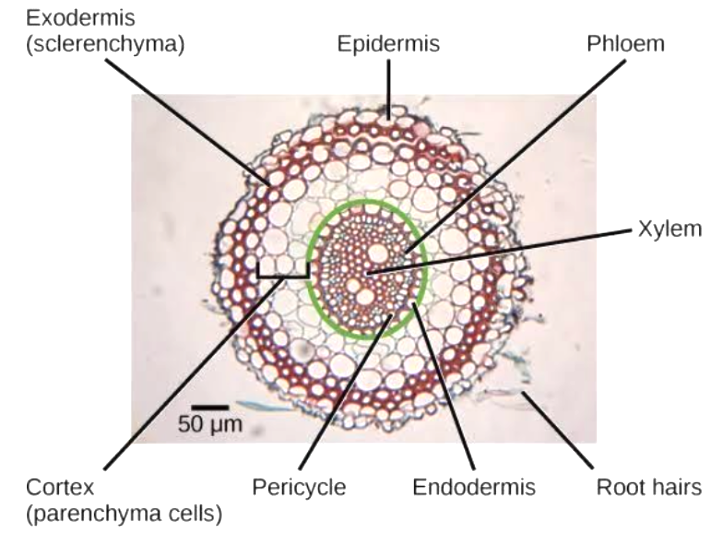
Below the epidermis lies the cortex, composed of loosely packed parenchyma cells with intercellular spaces. This region helps in the storage and transport of water and minerals.
Endodermis
The innermost layer of the cortex is characterised by barrel-shaped, tightly packed cells. It regulates the flow of substances into the vascular cylinder.
Pericycle
A few layers of thick-walled parenchyma lie immediately inside the endodermis. In dicots, the pericycle is the origin of lateral roots and contributes to secondary growth.
Vascular Bundle
In most dicot roots, there are two to four xylem and phloem strands arranged in a radial pattern.
The xylem and phloem are separated by parenchymatous conjunctive tissue. During secondary growth, a cambium develops between the xylem and phloem, leading to an increase in root girth.
Pith
In many dicot roots, the pith at the centre may be small or inconspicuous.
Monocot Root
Root System
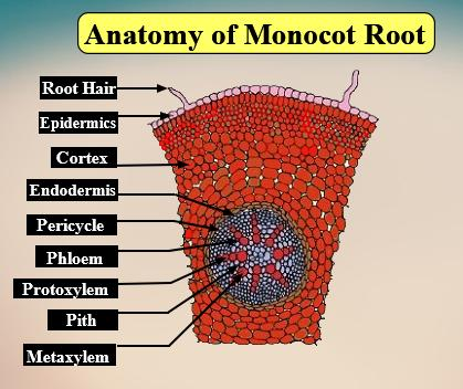
Monocots typically have an adventitious root system. Many thin roots emerge from the stem base or nodes close to the soil surface.
Epidermis and Cortex
The structure of dicot and monocot root, stem and leaf can appear quite similar in the outermost layers. In monocot roots, the epidermis is followed by a multilayered cortex of parenchyma cells.
Endodermis and Pericycle
The endodermis is well-defined, followed by a pericycle. However, secondary growth is generally absent in monocot roots, so the pericycle does not form a vascular cambium as in dicots.
Vascular Bundles
In monocots, the number of xylem strands is usually six or more (polyarch condition). Xylem and phloem alternate with each other in a ring-like arrangement.
Pith
Monocots often have a large and distinct pith in the centre of the root.
No Secondary Growth
Unlike dicots, monocots do not exhibit secondary growth in their roots.
Dicot Stem
A dicot stem is usually solid and exhibits distinct regions when viewed in cross-section:
Epidermis
The outermost protective layer is often covered by a thin cuticle. Epidermal cells may bear multicellular hairs (trichomes) and a few stomata.
Cortex
The cortex is typically divided into three sub-zones:
Hypodermis: Often made of collenchymatous cells, providing mechanical support.
Middle Cortical Layers: Composed of parenchyma with intercellular spaces for storage and transport.
Endodermis: The innermost boundary of the cortex. Also called the starch sheath in many dicots.
Pericycle
Situated beneath the endodermis. In stems, it may include patches of sclerenchyma.
Vascular Bundles
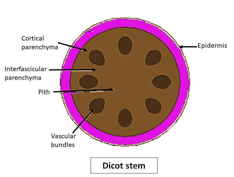
In a typical young dicot stem, vascular bundles are arranged in a ring. This arrangement is often referred to as a ring or circular pattern.
Each vascular bundle is conjoint (xylem and phloem together), open (a cambium present between xylem and phloem), and has an endarch protoxylem (protoxylem towards the centre).
This cambium is responsible for secondary growth, leading to the formation of annual rings.
Pith
The central region of the stem is composed of parenchyma cells that store nutrients and water.
Monocot Stem
A monocot stem is usually hollow or fibrous, and it does not undergo significant secondary growth:
Epidermis
A single-layered covering with a thick cuticle.
Hypodermis
Often consists of sclerenchyma, making the stem firm.
Vascular Bundles
The vascular bundles are numerous and scattered throughout the ground tissue (which includes both cortex and pith together). This scattered arrangement is a key feature in monocot vs dicot comparisons.
Vascular bundles are conjoint (xylem and phloem together) and closed (no cambium present), so there is no secondary growth.
Phloem parenchyma is generally absent.
Ground Tissue
Unlike dicots, monocots often lack a well-demarcated cortex and pith. Instead, they have a uniform ground tissue made up of parenchymatous cells.
Studying the anatomy of the monocot stem is crucial to understanding how monocots support themselves without secondary thickening.
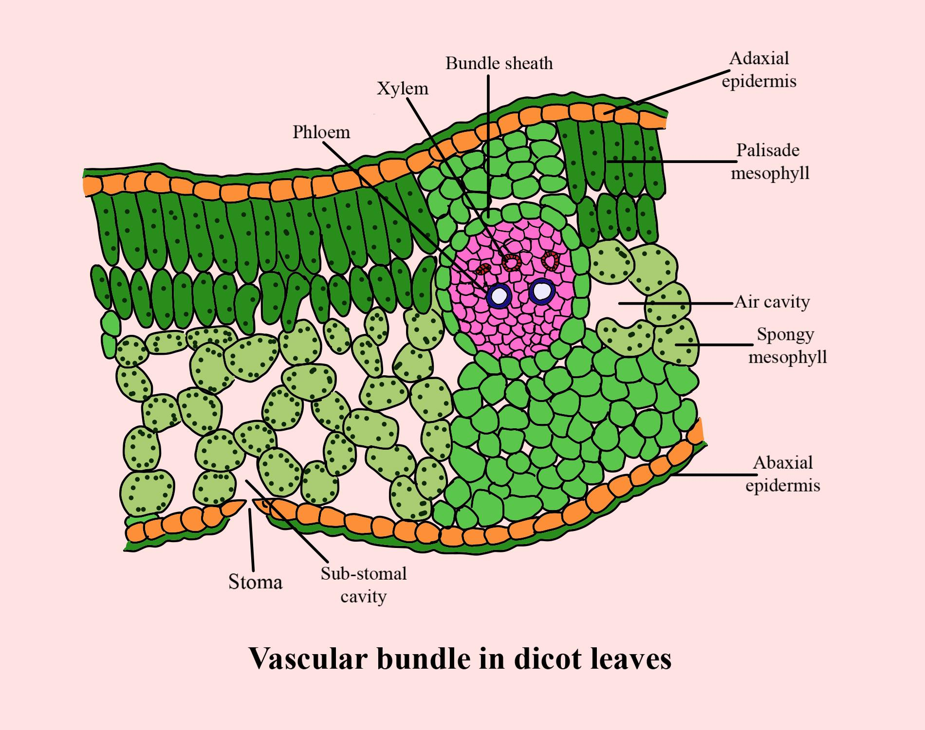
Dicot Leaf
Dicot leaves typically show reticulate (net-like) venation and can be divided into the following layers:
Epidermis
The upper (adaxial) and lower (abaxial) epidermal layers are usually covered by a cuticle.
Stomata may be present on both surfaces, though they are generally more abundant on the lower surface. Some dicot leaves may have stomata only on the lower surface.
Mesophyll
Consists of palisade parenchyma (elongated cells rich in chloroplasts, arranged just under the upper epidermis) and spongy parenchyma (loosely arranged cells with air spaces near the lower epidermis).
The mesophyll is the primary site of photosynthesis.
Vascular Bundles
Form the veins and midrib of the leaf, surrounded by a bundle sheath of parenchyma cells.
The vein arrangement (reticulate venation) distinguishes it from parallel venation in monocots.
Monocot Leaf
Monocot leaves are characterised by parallel venation:
Epidermis
Both upper (adaxial) and lower (abaxial) epidermal layers bear stomata, often arranged in rows.
Mesophyll
Typically, it is not differentiated into palisade and spongy parenchyma. The mesophyll is homogeneous.
Bulliform Cells
Large, thin-walled cells on the adaxial side help in leaf folding or rolling to reduce water loss during stress.
Vascular Bundles
Arranged parallel to each other, reflecting the parallel venation pattern.
By comparing the structure of dicot and monocot root, stem and leaf, you can see how these two groups differ in external morphology and internal arrangement of tissues.
Additional Differences
Below is a concise overview of monocot vs dicot differences for roots, stems, and leaves:
Root: Taproot in dicots vs adventitious roots in monocots.
Stem: Vascular bundles in a ring (dicots) vs scattered bundles (monocots).
Leaf: Reticulate venation (dicots) vs parallel venation (monocots).
Quick Quiz
Test your knowledge of the anatomy of dicotyledonous and monocotyledonous plants with these short questions. Answers are provided at the end.
1. Which layer in the roots is responsible for regulating the entry of water and minerals into the vascular bundle?
a) Cortex
b) Endodermis
c) Pericycle
d) Epidermis
2. What is the typical arrangement of vascular bundles in a dicot stem?
a) Scattered in the ground tissue
b) Concentric circles without cambium
c) Ring arrangement with cambium
d) Randomly placed with phloem parenchyma
3. In which type of leaf (dicot or monocot) are bulliform cells found?
a) Dicot leaves
b) Monocot leaves
4. Which type of stem usually has open vascular bundles?
a) Dicot stem
b) Monocot stem
Quiz Answers
b) Endodermis
c) Ring arrangement with cambium
b) Monocot leaves
a) Dicot stem
Related Topics


FAQs on Monocot and Dicot Anatomy: Roots, Stems, and Leaves Simplified
1. What are the main anatomical differences between a monocot and a dicot stem?
The primary differences in stem anatomy are found in the vascular bundles. In a dicot stem, vascular bundles are arranged in a ring and are 'open' (containing cambium), allowing for secondary growth. In a monocot stem, the vascular bundles are scattered throughout the ground tissue and are 'closed' (lacking cambium), so they do not undergo secondary growth.
2. How can you tell a monocot root from a dicot root by looking at its internal structure?
You can distinguish them by their vascular cylinders. A dicot root typically has a small number of xylem bundles (usually two to four, called tetrarch) and a less developed pith. In contrast, a monocot root has many more xylem bundles (usually six or more, called polyarch) and a large, well-developed pith at the center.
3. What is the difference between the leaf venation in monocots and dicots?
The pattern of veins in a leaf is a key identifier. Monocot leaves show parallel venation, where the veins run parallel to each other along the length of the leaf. Dicot leaves exhibit reticulate venation, where the veins form a branching, net-like pattern.
4. Why don't monocot plants typically show secondary growth (like getting wider)?
Monocots lack a specific tissue layer called the vascular cambium in their stems and roots. This cambium is responsible for producing new layers of xylem and phloem, which leads to an increase in girth or thickness (secondary growth). Since monocots have 'closed' vascular bundles without this cambium, they are generally unable to grow wider over time.
5. What is the main function of bulliform cells in some monocot leaves?
Bulliform cells are large, empty, colourless cells found on the upper surface of the leaves of certain monocots, like grasses. Their main function is to help regulate water loss. When water is plentiful, these cells are turgid and keep the leaf flat. During water scarcity, they lose water and become flaccid, causing the leaf to roll or fold inwards, which reduces the exposed surface area and minimises transpiration.
6. What does it mean for a vascular bundle to be 'open' or 'closed'?
This term describes the potential for growth. An 'open' vascular bundle, found in dicots, has a strip of cambium between the xylem and phloem, allowing it to produce more vascular tissues and grow. A 'closed' vascular bundle, found in monocots, does not have this cambium layer, so its growth potential is limited.
7. How does the mesophyll tissue differ between a monocot and a dicot leaf?
In a typical dicot leaf, the mesophyll tissue is differentiated into two layers: an upper palisade parenchyma (with elongated cells for photosynthesis) and a lower spongy parenchyma (with irregular cells and air spaces for gas exchange). In a monocot leaf, the mesophyll is usually not differentiated and consists of only spongy parenchyma.
8. How does the arrangement of vascular bundles affect the overall structure of monocot versus dicot plants?
The arrangement has a major impact on their form and strength. In dicots, the ring-like arrangement of vascular bundles (which later form a continuous cylinder with secondary growth) provides great structural support, allowing them to grow into large trees. In monocots, the scattered vascular bundles within a softer ground tissue do not provide the same kind of support for massive increases in width, which is why most monocots are herbaceous (non-woody), like grasses, corn, and lilies.


