

Labeled Structure and Functions of Mitochondria for Exams
Mitochondria play a vital role in the life of eukaryotic cells. Known popularly as the powerhouses of the cell, these rod-shaped organelles generate adenosine triphosphate (ATP), the main energy currency. They also take part in processes like cellular respiration, cell growth, and many other essential activities. Understanding the structure, and function of mitochondria can help you learn this crucial topic in biology.
What are Mitochondria?
Mitochondria are double-membraned, rod-shaped organelles found in the cytoplasm of most eukaryotic cells. The singular term ‘mitochondrion’ comes from Greek words meaning “thread” and “granule.” These organelles were first described by the German pathologist Richard Altmann in 1890.
Key Points
They are present in both plant cells and animal cells.
They are crucial for cellular respiration, differentiation, cell signalling, and controlling the cell cycle.
They have their genetic material (mitochondrial DNA) and ribosomes.
They produce ATP, which powers various biological processes.
Structure of Mitochondria
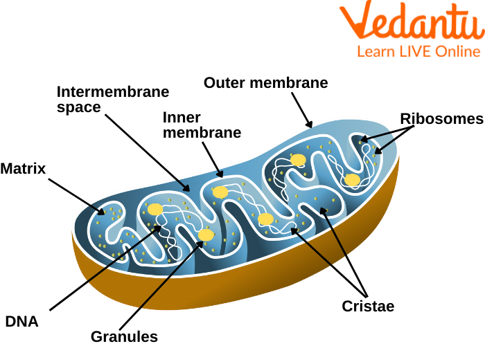
Matrix
The matrix is the viscous fluid enclosed by the inner membrane.
It contains enzymes for oxidative phosphorylation, ribosomes, mitochondrial DNA, ions, and other molecules.
It plays a key role in the Krebs cycle and in generating molecules that feed into ATP production.
Cristae
Cristae are the folds of the inner membrane that project into the matrix.
These folds greatly increase the surface area, enabling enhanced ATP production during cellular respiration.
Ribosomes
Mitochondrial ribosomes (mitoribosomes) synthesise some of the proteins needed by mitochondria.
They translate specific mRNAs encoded by the mitochondrial DNA.
Inner Membrane
The inner membrane contains several unique transporter proteins.
It is selectively permeable, which means it carefully regulates molecules entering or leaving the mitochondrial matrix.
Outer Membrane
The outer membrane contains proteins called porins, forming channels that allow smaller molecules to pass into the intermembrane space.
It also houses enzymes that help in various metabolic and signalling functions.
Intermembrane Space
This is the region between the inner and outer membranes.
It can be further divided into the intra-cristae space and the lumen, which are separated by narrow openings known as cristae junctions (10 to 40 nm in diameter).
It is important in protein transport, protein modification, and in regulating apoptosis (programmed cell death).
Function of Mitochondria
ATP Production: Mitochondria generate ATP through oxidative phosphorylation, a process that occurs along the inner membrane.
Cellular Respiration: They utilise oxygen to convert nutrients into energy.
Metabolic Activities: Mitochondria are involved in various metabolic pathways, including the Krebs cycle.
Cell Signalling: They help regulate calcium levels and influence signalling cascades that control cell growth.
Apoptosis: Mitochondria release factors that initiate programmed cell death, maintaining tissue health by removing damaged cells.
These roles underscore why these organelles are often called the “powerhouses” of the cell.
Quiz on Mitochondria
Test your understanding with these simple questions:
1. Which membrane of the mitochondria is folded to form cristae?
a) Outer membrane
b) Inner membrane
c) Intermembrane space
d) Matrix
Answer: b) Inner membrane
2. What is the main function of mitochondria?
a) Production of proteins
b) Storage of genetic material
c) ATP production
d) Cell wall formation
Answer: c) ATP production
3. Who first described mitochondria?
a) Gregor Mendel
b) Richard Altmann
c) Louis Pasteur
d) James Watson
Answer: b) Richard Altmann
Related Topics


FAQs on Diagram of Mitochondria with Detailed Structure Explanation
1. What is a diagram of mitochondria?
A diagram of mitochondria is a labeled representation that shows the structure and parts of a mitochondrion in detail. It typically includes the following components:
- Outer membrane
- Inner membrane
- Cristae (folds of the inner membrane)
- Matrix
- Intermembrane space
- Mitochondrial DNA and ribosomes
2. What are the main parts labeled in a mitochondria diagram?
The main parts labeled in a mitochondria diagram are the outer membrane, inner membrane, cristae, matrix, and intermembrane space. These parts include:
- Outer membrane – smooth boundary covering the organelle
- Inner membrane – folded membrane containing transport proteins
- Cristae – inward folds that increase surface area
- Matrix – inner fluid containing enzymes, mitochondrial DNA, and ribosomes
- Intermembrane space – space between the two membranes
3. What is the function of mitochondria in a cell?
The main function of mitochondria is to produce energy in the form of ATP (adenosine triphosphate) through cellular respiration. This occurs in stages:
- Glycolysis (in cytoplasm, linked to mitochondria)
- Krebs cycle (in the matrix)
- Electron transport chain (on the inner membrane)
4. What is the role of cristae in the mitochondria diagram?
The cristae increase the surface area of the inner membrane to enhance ATP production. They:
- Contain proteins of the electron transport chain
- Hold ATP synthase enzymes
- Allow more space for oxidative phosphorylation
5. What is the difference between the outer and inner membrane of mitochondria?
The outer membrane is smooth and permeable to small molecules, while the inner membrane is highly folded and selectively permeable. Key differences include:
- Outer membrane – contains porin proteins and forms the boundary
- Inner membrane – contains transport proteins and the electron transport chain
- The inner membrane forms cristae
6. What is the matrix in a mitochondria diagram?
The mitochondrial matrix is the innermost fluid-filled space enclosed by the inner membrane. It contains:
- Enzymes for the Krebs cycle
- Mitochondrial DNA (mtDNA)
- 70S ribosomes
- RNA and metabolic intermediates
7. Why do mitochondria have their own DNA?
Mitochondria have their own DNA because they originated from ancient bacteria through the endosymbiotic theory. Their mitochondrial DNA (mtDNA):
- Is circular like bacterial DNA
- Codes for some mitochondrial proteins
- Replicates independently of nuclear DNA
8. How does a mitochondrion produce ATP step by step?
A mitochondrion produces ATP through cellular respiration in a series of coordinated steps. These steps include:
- Pyruvate oxidation in the matrix
- Krebs cycle producing NADH and FADH₂
- Electron transport chain transferring electrons along the inner membrane
- Chemiosmosis where ATP synthase forms ATP
9. Are mitochondria present in plant and animal cells?
Yes, mitochondria are present in both plant and animal cells as well as most eukaryotic organisms. In:
- Animal cells – they provide energy for movement and metabolism
- Plant cells – they produce ATP in addition to energy made in chloroplasts
10. Why are mitochondria called the powerhouse of the cell?
Mitochondria are called the powerhouse of the cell because they generate most of the cell’s ATP through cellular respiration. They:
- Break down glucose and other molecules
- Convert energy into usable ATP
- Support essential cellular processes like active transport and muscle contraction


