

What Are the Structure and Functions of the Alimentary Canal?
The alimentary canal anatomy is central to understanding how food is processed in the human body. This long, muscular tube starts at the mouth and ends at the anus, ensuring that nutrients are properly digested, absorbed, and wastes eliminated. Studying this anatomy helps us learn about essential processes like digestion, and is important for students, healthcare professionals, and anyone interested in human biology.
What is Alimentary Canal Anatomy?
Alimentary canal anatomy refers to the structure and organisation of the continuous passage extending from the mouth to the anus through which food travels. This system, also called the digestive or gastrointestinal tract, includes important organs like the mouth, pharynx, esophagus, stomach, small intestine, and large intestine. It is critical for transforming food into energy and nutrients required for life.
Key Parts of the Alimentary Canal Anatomy
1. Mouth (Oral Cavity)
The mouth is the beginning of the alimentary canal anatomy. It functions in the ingestion and initial mechanical and chemical breakdown of food. Chewing with teeth (mastication) and mixing food with saliva softens and prepares it for swallowing. The mouth also contains structures like the tongue, hard and soft palate, uvula, and three pairs of salivary glands (parotid, submandibular, and sublingual). Saliva not only lubricates but introduces enzymes like amylase, beginning carbohydrate digestion.
- Teeth cut, tear, and grind food into small pieces for easier swallowing.
- The tongue shapes chewed food into a bolus, making it easier to swallow.
- Salivary amylase starts the chemical breakdown of starch into simpler sugars.
- The oral cavity supports speech and also plays a role in breathing.
2. Pharynx (Throat)
The pharynx is a muscular tube that connects the mouth to the esophagus and the larynx. It is divided into three regions: nasopharynx, oropharynx, and laryngopharynx. Its main roles include channelling food from the mouth to the esophagus and air to the lungs, making it vital for both digestion and respiration.
- The oropharynx lies behind the oral cavity, receiving food after it is swallowed.
- The epiglottis, a flap of tissue in the pharynx, prevents food from entering the windpipe during swallowing.
- Lymphoid tissues like tonsils, found in the pharynx, are part of the immune system.
3. Esophagus
The esophagus is a long, muscular tube that runs from the pharynx to the stomach. It acts as a passageway, moving food using coordinated muscular contractions called peristalsis. The esophageal lining also secretes mucus for smooth passage of food. The esophagus passes through the diaphragm to join the stomach, and the junction is regulated by the lower esophageal sphincter, preventing the backflow of stomach contents.
- Peristalsis pushes the bolus (chewed food) towards the stomach.
- The esophageal sphincters (upper and lower) control entry and exit of materials.
- Mucus produced by the lining aids in lubrication and easy movement.
4. Stomach
The stomach is a muscular, J-shaped organ that acts as a temporary storage and mixing chamber. Food mixes with gastric juices, which contain enzymes and hydrochloric acid. This process turns the food into a semi-liquid substance called chyme. The stomach is key in starting protein digestion. You can read more about stomach anatomy and related processes on our stomach page.
- Gastric glands secrete pepsinogen and hydrochloric acid for protein breakdown.
- The stomach churns, mixes, and liquefies food.
- Sphincters at both ends (cardiac and pyloric) control entry and exit.
5. Small Intestine
The small intestine is the longest part of the alimentary canal anatomy. It is responsible for the majority of digestion and nutrient absorption. It consists of three sections: duodenum, jejunum, and ileum. Enzymes from the pancreas and bile from the liver further break down food. The inner surface is lined with villi and microvilli to maximize absorption. Learn more about absorption and function in the Small Intestine notes.
- Duodenum is the first and shortest section, receiving secretions from pancreas and liver.
- Jejunum and ileum are specialized for digestion and absorption.
- Intestinal villi increase the surface area for efficient nutrient absorption.
6. Large Intestine
The large intestine finishes the absorption of water and salts, forms stool, and eliminates waste. It includes the cecum, colon, rectum, and anus. Helpful gut bacteria further digest some substances and produce vitamins. Waste is compacted as it moves towards the anus for excretion.
- Removes water and electrolytes from undigested food.
- Hosts beneficial bacteria involved in fermentation and vitamin production.
- Stores stool before it is expelled through the anus.
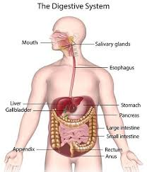
Alimentary Canal Anatomy Diagram
A well-labelled diagram is essential for understanding the layout and organ placement of the alimentary canal anatomy. You can also explore labelled diagrams in Vedantu's dedicated digestive system diagram section for visual reference.
Alimentary Canal Anatomy Definition
The alimentary canal is defined as a continuous passage in the human body starting from the mouth and ending at the anus, through which food passes and is digested. It is the core component of the digestive system, working closely with accessory digestive organs.
Functions of the Alimentary Canal
The main roles of the alimentary canal anatomy include breaking down complex food substances, absorbing useful nutrients, and removing waste. It achieves these functions through specialized organs and coordinated muscular movements.
- Ingestion: Taking in food through the mouth.
- Mechanical and Chemical Digestion: Breaking down food into smaller, absorbable components.
- Absorption: Transporting nutrients into the bloodstream through the intestinal wall.
- Assimilation: Using absorbed nutrients for growth, energy, and repair.
- Egestion: Removing undigested and unabsorbed food as waste.
Alimentary Canal Anatomy Examples
Humans share similar alimentary canal anatomy with many animals. For example, the digestive tracts of cows, earthworms, and pigs have sections performing comparable functions—ingestion, digestion, absorption, and egestion. However, species may differ in the length and specialization of certain regions, such as the highly compartmentalised stomach in ruminants or a simple sac-like digestive tract in some invertebrates.
- Human digestive system
- Cow (ruminant) digestive tract
- Earthworm alimentary canal
Short Notes on Alimentary Canal Anatomy
The alimentary canal anatomy consists of a complex tube divided into mouth, pharynx, esophagus, stomach, small intestine, and large intestine. Each segment has distinct roles in the overall process of digestion and absorption, supported by accessory organs such as the liver and pancreas. A detailed understanding of this system is vital for grasping topics in life science and food science.
Alimentary Canal Anatomy Questions and MCQs
Alimentary canal anatomy is a popular subject in class 12 and competitive biology exams. Students often encounter a variety of MCQs and short notes questions regarding the structure, function, and characteristics of the digestive tract. Practicing these can reinforce understanding and improve exam performance.
- Which organ is responsible for the initial breakdown of starch?
- List the main parts of the alimentary canal in sequence.
- What is the function of the epiglottis?
Real-World Relevance & Applications
Understanding alimentary canal anatomy helps medical professionals diagnose and treat digestive disorders. This knowledge is also relevant in developing nutrition plans, improving food processing, and teaching healthy eating habits. The system’s health can be influenced by the environment, food choices, and medical conditions like diabetes or vitamin deficiencies. Explore related topics like nutrients and their functions and digestion definitions for broader context.
Alimentary Canal Anatomy PPT, Notes, and Study Guides
Students looking for further resources, such as alimentary canal anatomy PPTs or detailed notes, can find excellent study materials on Vedantu. These resources break down complex concepts, offer diagrams for easy visualization, and include practice questions for effective exam preparation.
Mastering alimentary canal anatomy provides a foundation for learning about human health, disease prevention, and nutrition. This knowledge connects students to broader topics within biology and everyday life. For more support, explore other learning resources and biology notes available on Vedantu’s platform.
In summary, alimentary canal anatomy covers the detailed study of the organs and functions involved in human digestion, starting from the mouth to the anus. It emphasizes the importance of each segment in breaking down food, nutrient absorption, and waste removal. Understanding this topic supports academic excellence and promotes lifelong health.


FAQs on Understanding Alimentary Canal Anatomy
1. What is the alimentary canal and what are its main functions?
The alimentary canal is a continuous muscular tube that runs from the mouth to the anus and is responsible for digestion and absorption of food. It performs the following main functions:
- Ingestion: Taking in food through the mouth.
- Digestion: Breaking down food into smaller, absorbable molecules.
- Absorption: Nutrients pass from the digestive tract into the bloodstream.
- Elimination: Removal of undigested waste via the anus.
2. What are the parts of the human alimentary canal?
The human alimentary canal consists of several main parts that form the digestive tract:
- Mouth
- Pharynx
- Oesophagus
- Stomach
- Small intestine (duodenum, jejunum, ileum)
- Large intestine (colon, rectum)
- Anus
3. What is the function of the small intestine in the alimentary canal?
The small intestine is the main site for digestion and absorption of nutrients in the alimentary canal. It:
- Receives partially digested food (chyme) from the stomach
- Uses digestive enzymes and bile to break down carbohydrates, proteins, and fats
- Absorbs nutrients through its villi into the bloodstream
4. How does food move through the alimentary canal?
Food moves through the alimentary canal by a process called peristalsis, which involves:
- Rhythmic contractions of smooth muscles in the canal walls
- Pushing food and digestive contents from the mouth to the anus
- Ensuring continuous and controlled movement for effective digestion
5. What is the difference between alimentary canal and digestive system?
The alimentary canal is the hollow tube extending from mouth to anus, whereas the digestive system includes the alimentary canal and accessory organs (like liver, pancreas, and salivary glands) that aid digestion.
6. Name the digestive glands associated with the alimentary canal.
The digestive glands associated with the alimentary canal are:
- Salivary glands
- Liver
- Pancreas
- Gastric glands in the stomach
- Intestinal glands in the small and large intestine
7. What is the structure and function of the stomach in the alimentary canal?
The stomach is a muscular, J-shaped organ in the alimentary canal:
- Stores and mixes food with gastric juices
- Initiates protein digestion with the enzyme pepsin
- Regulates entry of food into small intestine as chyme
8. Which part of the alimentary canal is mainly responsible for water absorption?
The large intestine is mainly responsible for absorbing water from undigested food material, forming semi-solid feces for excretion.
9. List the steps of digestion as food passes through the alimentary canal.
Digestion in the alimentary canal occurs in a stepwise manner:
- Ingestion – Food is taken into the mouth.
- Mechanical breakdown – Chewing and churning break food into smaller pieces.
- Chemical digestion – Enzymes break down large molecules in the stomach and small intestine.
- Absorption – Nutrients transported across intestinal walls into the blood.
- Elimination – Waste expelled through the anus.
10. What are the accessory organs of the digestive system and their roles?
The accessory organs include the liver, pancreas, and gallbladder. Their roles:
- Liver: Produces bile for fat digestion.
- Pancreas: Secretes digestive enzymes and hormones.
- Gallbladder: Stores and releases bile into small intestine.
11. How long is the human alimentary canal?
The human alimentary canal is approximately 8-10 metres long in adults, stretching from the mouth to the anus.
12. Why is the small intestine so long compared to other parts of the alimentary canal?
The small intestine is very long (around 6 meters) to provide a large surface area for maximum absorption of nutrients, with millions of villi and microvilli increasing the contact area inside the intestine.


